
Protected: MRI Transitional Lumbosacral Anatomy Ilio Lumbar Ligament
There is no excerpt because this is a protected post.
There is no excerpt because this is a protected post.
CLICK HERE FOR OUR LATEST SPINE MRI MINI-FELLOWSHIPS Radiology Conference Malaysia Kuala Lumpur. This meeting is now over. If you would like to see photos from the event, please follow this link to our Facebook page KLSpinePhotos We have had a lot of requests to hold the Spine Workstation Workshop in Kuala Lumpur as well …
Spine and SIJ MRI Workstation Workshop Kuala Lumpur Read More »
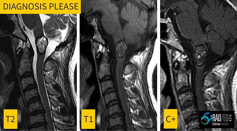
This is the first in a regular series of posts on cases of interest. Incidental finding in a 40yo male. Image above. ANSWER Cavernomas of the spinal cord have a very specific appearance on MRI with the following features Lobulated well defined lesion mixed signal T1 and T2 No significant enhancement Low signal …
Radiology Education Diagnosis Please: Spinal cord lesion MRI Read More »

The 2016 ISMRM Annual Meeting is being held in Singapore, which is great news for us in this region. But is it worth going to? It depends…. I have been to a number of ISMRM meetings over the years and its a huge meeting, but the difficulty from a radiologist’s perspective, is that it …
PARS DEFECTS ON MRI Pars defects can be difficult to see on MRI. Lets look at how best to find them 1. WHAT IS THE PARS INTERARTICULARIS Bone that connects the superior and inferior articular process at each level. 2. WHAT DOES A NORMAL PARS INTERTICULARIS LOOK LIKE ON MRI A continuous piece of bone …
INTRO: Pars defects can be difficult to see on MRI. Here is an easy way to know if the slip/ spondylolistheisis you are seeing is due to a pars defect or is from degenerative facet disease. THE TIP: In Degenerative spondylolisthesis, the canal is always small. In spondylolisthesis due to a Pars Defect, the …
SPONDYLOLISTHESIS: PARS DEFECT OR DEGENERATION: HOW TO DIFFERENTIATE QUICKLY Read More »