SEO

hamstring tendon mri anatomy origin conjoined conjoint tendon origin tendinitis tendonosis rea

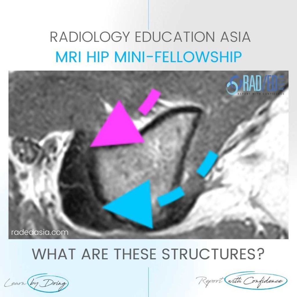
HIP MRI HAMSTRING CONJOINT TENDON ORIGIN ANATOMY

HIP MRI HAMSTRING CONJOINT TENDON ORIGIN ANATOMY DISCUSSION This is the hamstring tendon origin at ischial tuberosity. The Anterior tendon is SEMIMEMBRANOSUS (Pink arrow). The Posterior tendon is the CONJOINT TENDON (Blue arrow) consisting of: SEMITENDINOSUS BICEPS FEMORIS Both demonstrate  normal signal which is a uniform black signal on all sequences. HIP MRI HAMSTRING CONJOINT …

HIP MRI HAMSTRING CONJOINT TENDON ORIGIN ANATOMY Read More »

Imaging Rheumatology Radiology MRI Rheumatologists radedasia

IMAGING ARTHRITIS RHEUMATOLOGY ANKYLOSING SPONDYLOARTHRITIS MRI & RADIOLOGY

Home IMAGING ARTHRITIS RHEUMATOLOGY ANKYLOSING SPONDYLITIS RADIOLOGY FOR RHEUMATOLOGISTS & RADIOLOGISTS: LEARN RHEUMATOLOGY MRI RADIOLOGY ONLINE IMAGING ARTHRITIS RHEUMATOLOGY ANKYLOSING SPONDYLITIS RADIOLOGY & MRI  What to Look for, Where to Look and How to Best Report it. RHEUMATOLOGY AND RADIOLOGY: Welcome to our page on Imaging Arthritis, Rheumatology Ankylosing Spondylitis where all our Rheumatology and Arthritis imaging posts will be available. …

IMAGING ARTHRITIS RHEUMATOLOGY ANKYLOSING SPONDYLOARTHRITIS MRI & RADIOLOGY Read More »

MRI Spine Rheumatologist rheumatology Ankylosing Spondylitis Spondyloarthropathy radedasia

MRI SPINE RHEUMATOLOGY FOR RHEUMATOLOGISTS: HOW TO ASSESS ANKYLOSING SPONDYLITIS AND SPONDYLOARTHROPATHY PART 1

MRI SPINE FOR RHEUMATOLOGISTS & (RADIOLOGISTS) How to assess Ankylosing Spondylitis and Spondyloarthropathy Part 1: The Sequences you need to know. Rheumatologists should be able to assess a MRI Spine for changes of Ankylosing Spondylitis and Spondyloarthropathy. We have a 3 part series for Rheumatologists (and Radiologists 😀) on how to have a structure to assess …

MRI SPINE RHEUMATOLOGY FOR RHEUMATOLOGISTS: HOW TO ASSESS ANKYLOSING SPONDYLITIS AND SPONDYLOARTHROPATHY PART 1 Read More »

meniscus cyst radiology intrameniscal tear mri knee

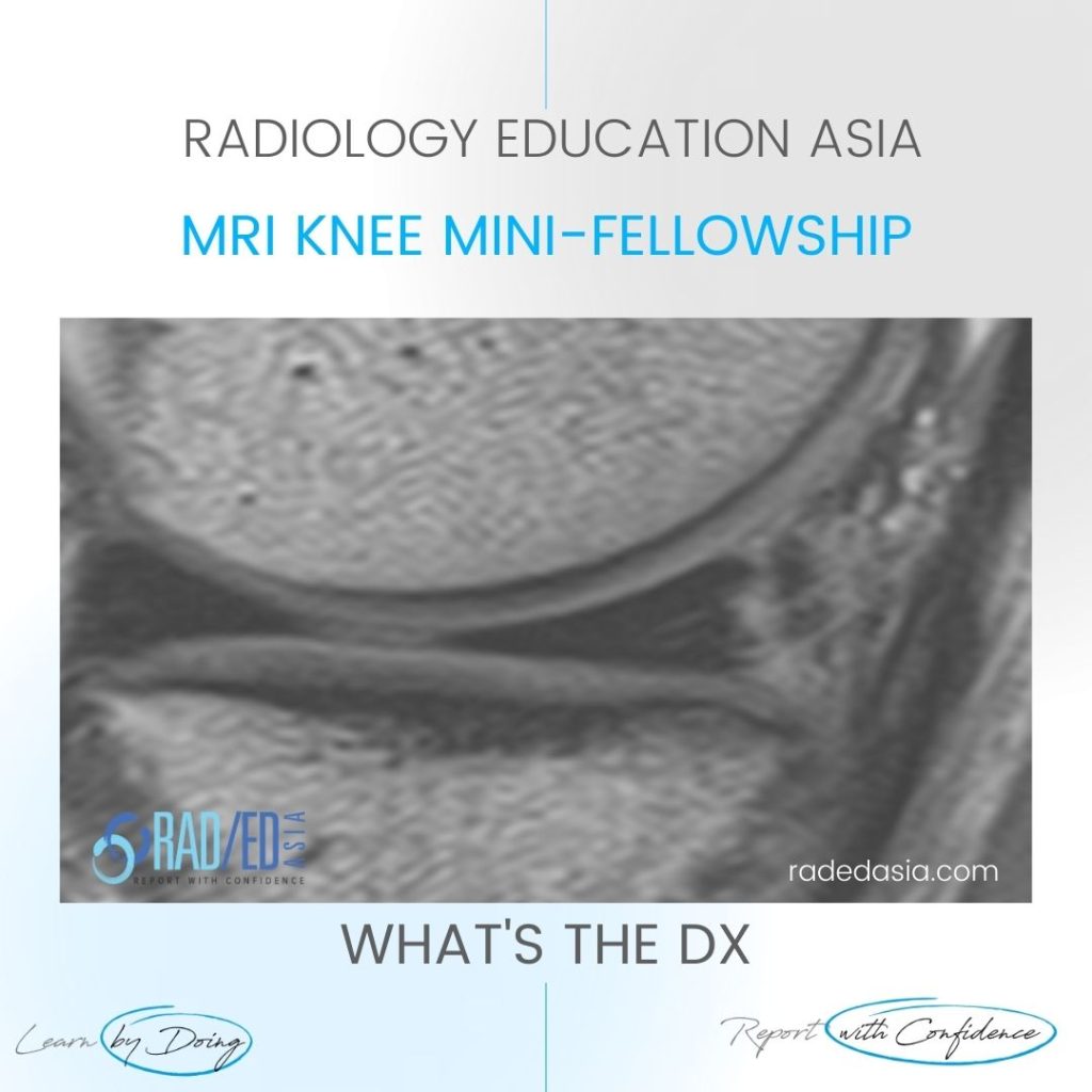
MENISCAL CYST RADIOLOGY INTRAMENISCAL TEAR MRI KNEE

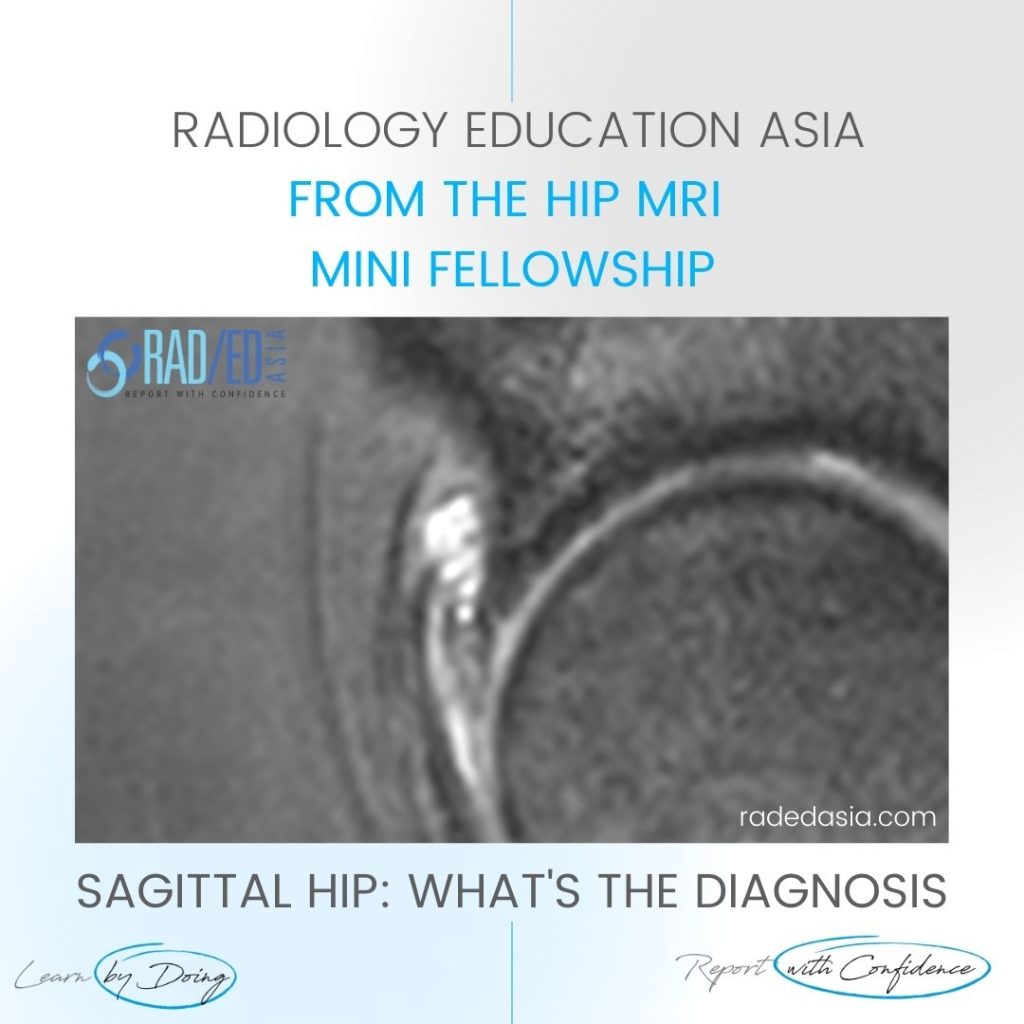
MENISCAL INTRAMENISCAL CYST MRI RADIOLOGY DISCUSSION INTRAMENISCAL CYST. Extensive intra meniscal cyst anterior horn. A tear is not seen extending to the articular margin. However we may not always see the extension on MRI. So if you see an intra-meniscal or parameniscal cyst these are always due to an underlying tear even if we don’t …

MENISCAL CYST RADIOLOGY INTRAMENISCAL TEAR MRI KNEE Read More »

biceps labrum anchor complex slap tear attachment insertion mri

SHOULDER MRI BICEPS LABRUM JUNCTION: Q&A FROM THE COURSE

We have had over 2000 questions answered in the MRI Mini Fellowships. From simple to complex and everything in between. Here are some of them. QUESTION: Q: Is it normal to see a fluid cleft between he biceps tendon and superior labrum? MOVE SLIDER ICON (ARROWS) LEFT TO SEE THE ANSWER ANSWER: A: Hi, Dr …

SHOULDER MRI BICEPS LABRUM JUNCTION: Q&A FROM THE COURSE Read More »

cord signal change spondylotic myelopathy mri

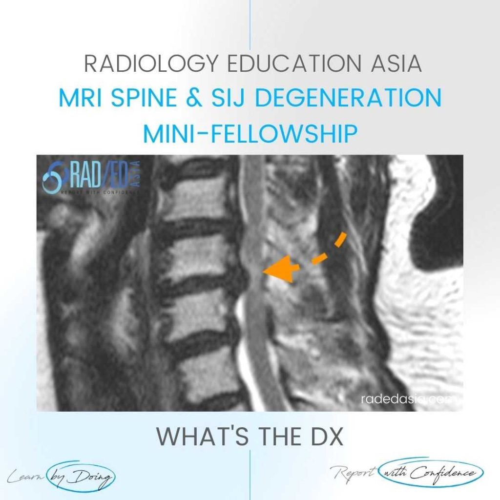
CORD SIGNAL CHANGE MRI SPONDYLOTIC MYELOPATHY

SPONDYLOTIC MYELOPATHY MRI RADIOLOGY DISCUSSION SPONDYLOTIC MYELOPATHY   Spondylotic Myelopathy is a term used when there is increased cord signal due to extrinsic compression.   Patients will often be symptomatic with cord related symptoms.   By using the term you are indicating that the patient’s symptoms can potentially be reversed by relieving the compression. WHAT …

CORD SIGNAL CHANGE MRI SPONDYLOTIC MYELOPATHY Read More »

rotator cuff tendinitis mri supraspinatus tendinosis bursitis radiology

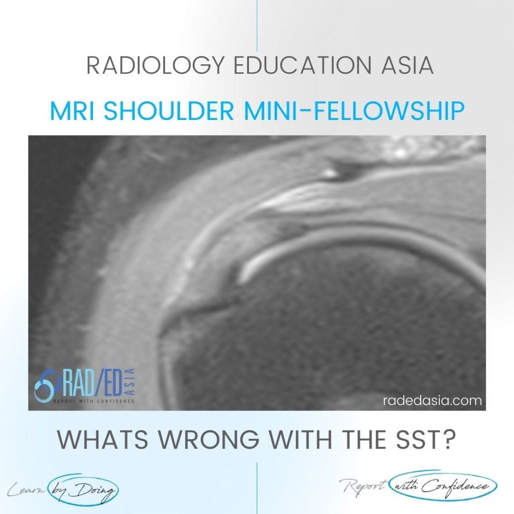
ROTATOR CUFF SUPRASPINATUS TENDINOSIS TENDINITIS MRI: WHAT’S WRONG WITH THE SST?

MOVE SLIDER ICON (ARROWS) LEFT TO SEE THE DIAGNOSIS WHAT ARE THE FINDINGS Diffuse Increased signal (intermediate) in the Supraspinatus Tendon. WHAT'S THE Dx SUPRASPINATUS TENDINITIS/TENDINOSIS.  There is diffuse increased signal in the supraspinatus tendon (Pink arrows). The signal increase is intermediate which is seen on tendinitis and not fluid type signal which is seen …

ROTATOR CUFF SUPRASPINATUS TENDINOSIS TENDINITIS MRI: WHAT’S WRONG WITH THE SST? Read More »

This site is intended for Medical Professionals only. Use of this site is governed by our Terms of Service and Privacy Statement which can be found by clicking on the links. Please accept before proceeding to the website.
error: Right Click not available.