SEO

msk webinar conference musculoskeletal mri

MSK WEBINAR STRESS FRACTURES ONLINE

MSK MRI WEBINAR: STRESS FRACTURES MSK WEBINAR IMAGING OF STRESS FRACTURES Join us for talks by Dr Ravi Padmanabhan and Prof Ichiro Sekaya on Imaging of Stress Fractures and The Relationship between Medial Tibial Osteophytes and Meniscal Extrusion The webinar is presented and organized by Fuji Film Singapore. MSK WEBINAR ON 6th FEBRUARY 2022, 4.30 …

MSK WEBINAR STRESS FRACTURES ONLINE Read More »

biceps tenosynovitis mri tendon sheath effusion tendinitis tendonitis radedasia

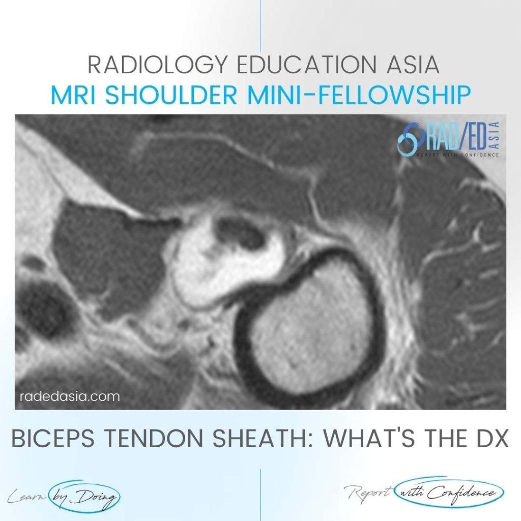
BICEPS TENDON SHEATH TENOSYNOVITIS EFFUSION MRI

BICEPS TENDON SHEATH TENOSYNOVITIS EFFUSION MRI DISCUSSION ON BICEPS TENOSYNOVITIS & TENDON SHEATH EFFUSION BICEPS TENDON SHEATH TENOSYNOVITIS   There can normally be a small amount of fluid in the biceps tendon sheath.   However in this case there is markedly increased fluid in the biceps tendon sheath in keeping with tenosynovitis.   Additionally there …

BICEPS TENDON SHEATH TENOSYNOVITIS EFFUSION MRI Read More »

tibialis posterior tendinopathy mri radiology radedasia

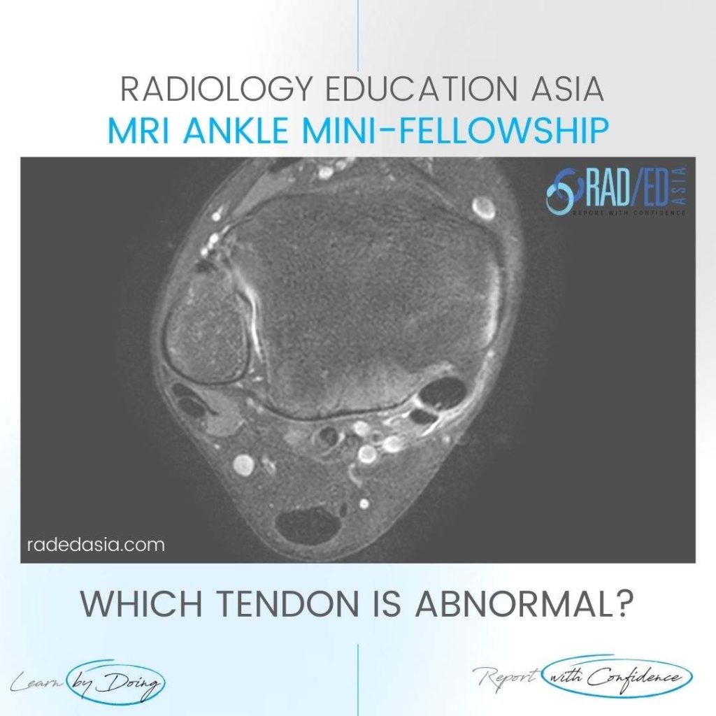
TIBIALIS POSTERIOR TENDINOPATHY MRI RADIOLOGY

TIBIALIS POSTERIOR TENDINOPATHY MRI RADIOLOGY DISCUSSION TIBIALIS POSTERIOR TENDINOPATHY MRI APPEARANCE Tibialis Posterior Tendinopathy on MRI can have a number of appearances.   One of the features is tendon enlargement with the signal remaining normal.   The normal tibialis posterior tendon should be less than twice the size of the adjacent Flexor Hallicus Longus (Green …

TIBIALIS POSTERIOR TENDINOPATHY MRI RADIOLOGY Read More »

synovitis mri ighl shoulder

MRI SYNOVITIS SPECIFIC SITES IN THE SHOULDER: THE INFERIOR GLENOHUMERAL LIGAMENT IGHL

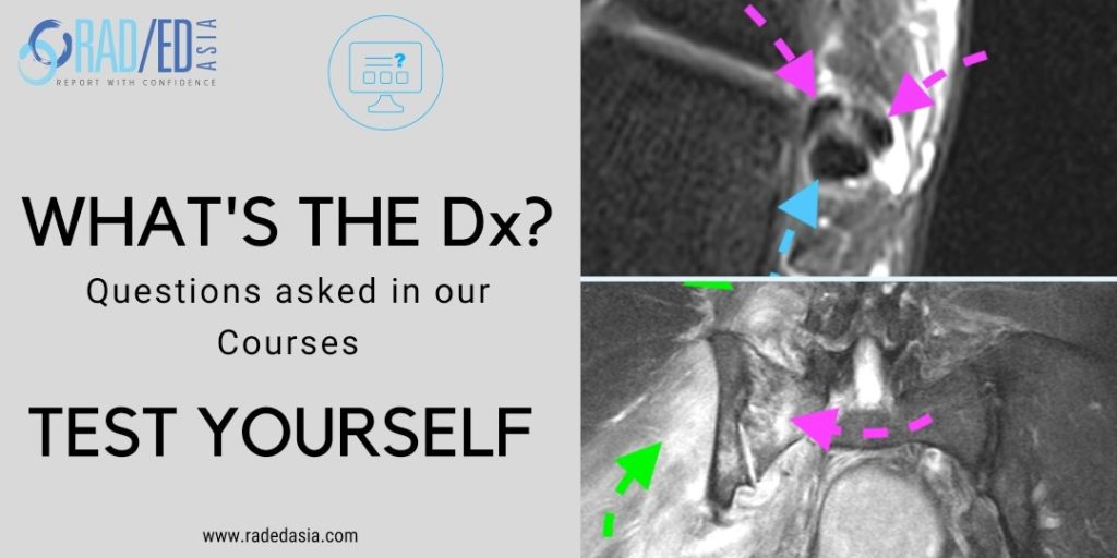
MRI Shoulder Synovitis Capsulitis: Inferior Glenohumeral Ligament IGHL Capsulo Synovitis MRI Capsulo Synovitis IGHL. At our MRI workshops we have an Open Mic policy. People are encouraged to interrupt and ask questions at anytime because its best to clear doubts at the time topics are being discussed rather than wait till the end or never …

MRI SYNOVITIS SPECIFIC SITES IN THE SHOULDER: THE INFERIOR GLENOHUMERAL LIGAMENT IGHL Read More »

bifurcate ligament radiology anterior mri anatomy

BIFURCATE LIGAMENT RADIOLOGY MRI ANATOMY ANKLE

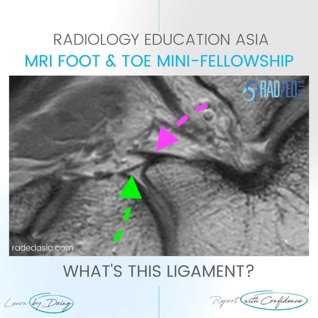
BIFURCATE LIGAMENT RADIOLOGY MRI ANATOMY ANKLE DISCUSSION ON BIFURCATE LIGAMENT This is the Bifurcate ligament.   It arises from the anterior process of the calcaneum (Yellow arrow).   Its has two components that attaches: To the Navicular (Pink arrow). To the Cuboid (Green Arrow). KEY POINT ON BIFURCATE LIGAMENT On MRI, the Bifurcate Ligament is …

BIFURCATE LIGAMENT RADIOLOGY MRI ANATOMY ANKLE Read More »

patella dislocation subluxation fracture hemarthrosis lipohemarthrosis radiology mri xray knee

PATELLA DISLOCATION SUBLUXATION FRACTURE HEMARTHROSIS LIPOHEMARTHROSIS MRI KNEE RADIOLOGY

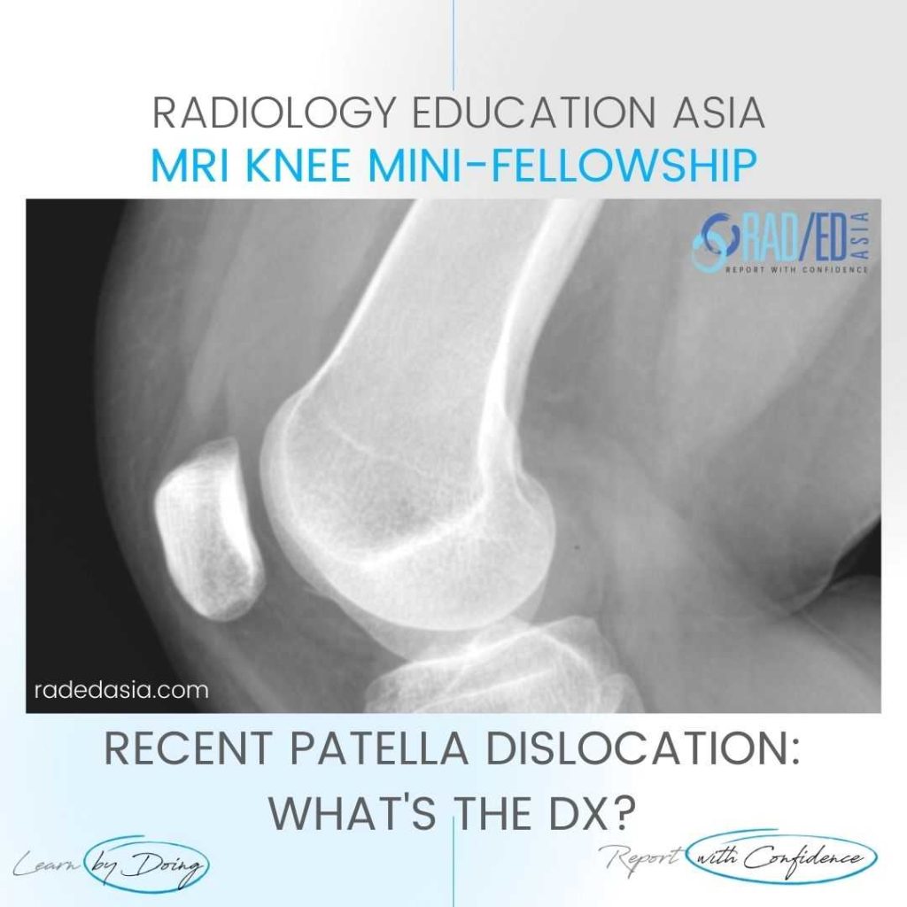
PATELLA DISLOCATION SUBLUXATION HEMARTHROSIS FRACTURE DISCUSSION ON PATELLA DISLOCATION SUBLUXATION HEMARTHROSIS FRACTURE The patient has had a recent patella dislocation and there is now a large suprapatellar effusion (on MRI this was a hemarthrosis).  On X-ray we can’t say defnitely that it’s a hemarthrosis but the presence of a fracture would indicate that it is …

PATELLA DISLOCATION SUBLUXATION FRACTURE HEMARTHROSIS LIPOHEMARTHROSIS MRI KNEE RADIOLOGY Read More »

retrocalcaneal bursa bursitis achilles tendon mri radiology radedasia

RETROCALCANEAL BURSITIS RADIOLOGY MRI ANKLE

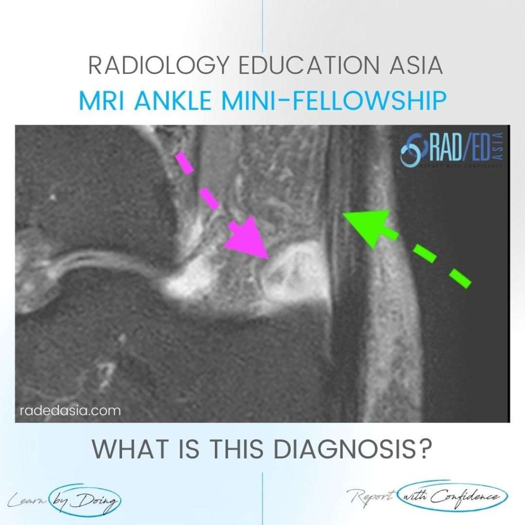
RETROCALCANEAL BURSITIS RADIOLOGY MRI WHAT'S THE Dx RETROCALCANEAL BURSITIS. ACHILLES TENDONOSIS. DISCUSSION The retrocalcaneal bursa lies anterior and deep to the Achilles tendon postero superior to the calcaneum. The normal bursa has minimal fluid so on MRI you may see nothing or just a thin amount of fluid without synovial thickening. For retrocalcaneal bursitis on …

RETROCALCANEAL BURSITIS RADIOLOGY MRI ANKLE Read More »

spine mri ankylosing spondylitis spondyloarthritis SpA rheumatologist rheumatology radiology

MRI SPINE ANKYLOSING SPONDYLITIS SpA SPONDYLOARTHRITIS FOR RHEUMATOLOGISTS: HOW TO ASSESS: PART 2 WHAT DOES THE SIGNAL MEAN

MRI SPINE FOR RHEUMATOLOGISTS & (RADIOLOGISTS) In this video below we look at how to assess a MRI Spine Ankylosing Spondylitis & Spondyloarthritis (SpA ) for Rheumatologists (and Radiologists ) and look at the SIGNAL CHANGES YOU NEED TO KNOW AND WHAT  THEY MEAN. This is the 2nd part of a 3 part series on …

MRI SPINE ANKYLOSING SPONDYLITIS SpA SPONDYLOARTHRITIS FOR RHEUMATOLOGISTS: HOW TO ASSESS: PART 2 WHAT DOES THE SIGNAL MEAN Read More »

This site is intended for Medical Professionals only. Use of this site is governed by our Terms of Service and Privacy Statement which can be found by clicking on the links. Please accept before proceeding to the website.
error: Right Click not available.