SEO

scapula normal variant keats shoulder mri radiology radedasia

NORMAL SCAPULA VARIANT SHOULDER MRI RADIOLOGY (VIDEO)

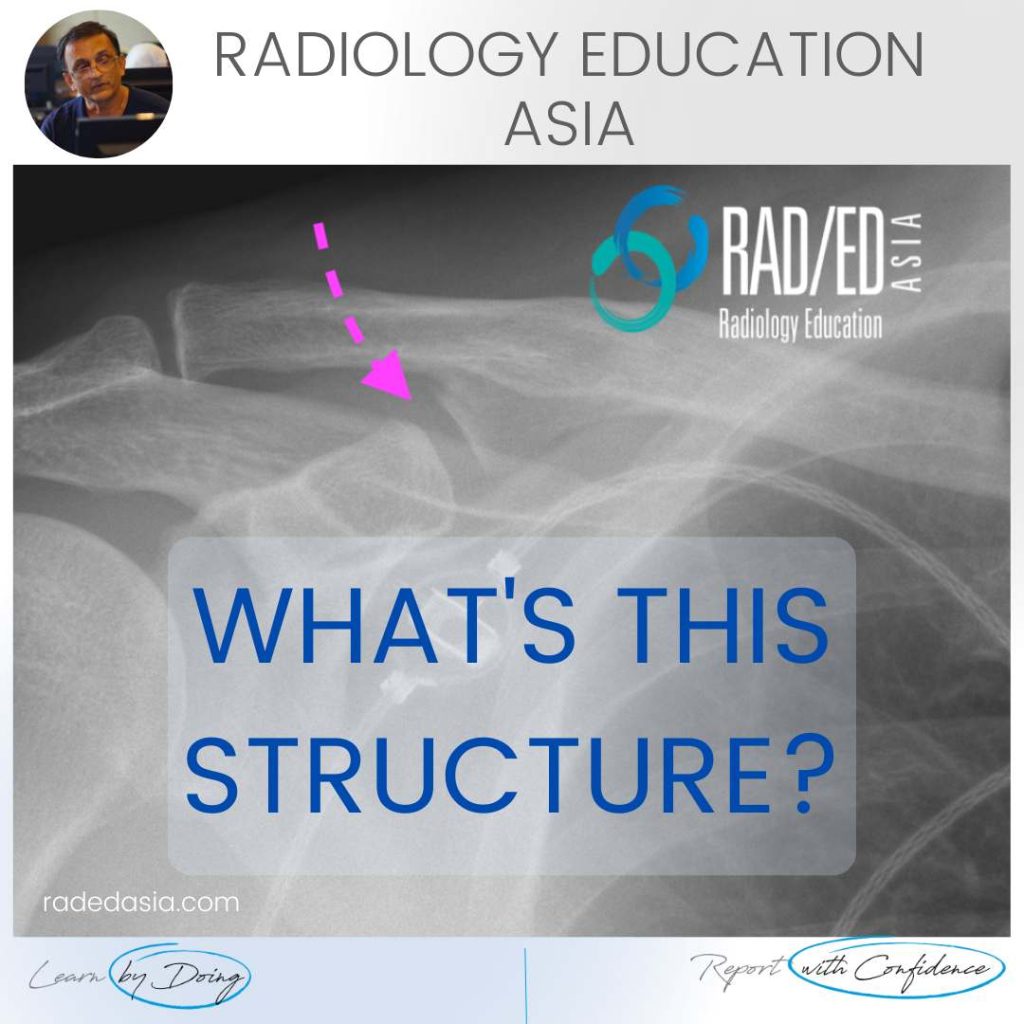
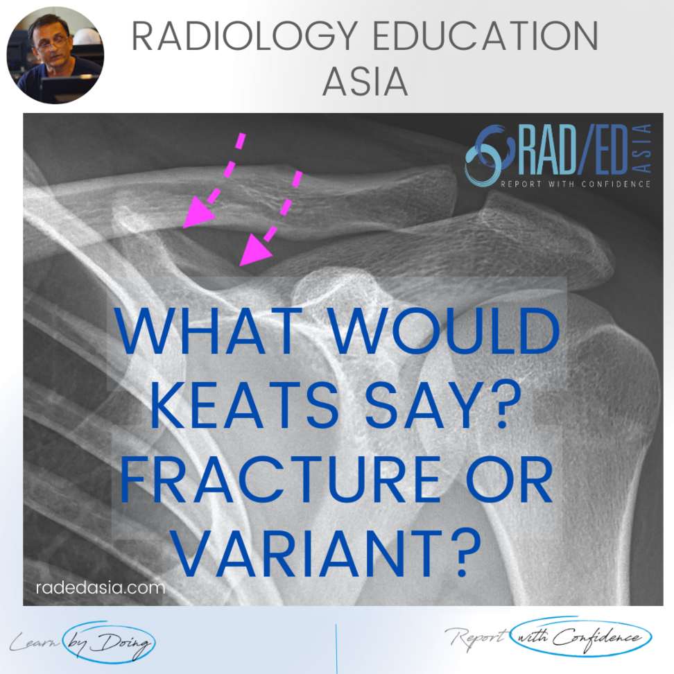
NORMAL SCAPULA VARIANT: CLASP LIKE SUPERIOR MARGIN FINDINGS Well corticated bridge of bone (Pink arrows) that lies on the superior margin of the scapula with a bone defect inferior to it. DISCUSSION This is a normal variant. Its described in Keats as a Clasp Like scapular margin that’s a result of bone being developmentally absent …

NORMAL SCAPULA VARIANT SHOULDER MRI RADIOLOGY (VIDEO) Read More »

popliteomeniscal fasicle mri radiology tear anatomy lateral knee radedasia

POPLITEOPMENISCAL FASICLE MRI RADIOLOGY KNEE (VIDEO)

POPLITEOMENISCAL FASICLE MRI DISCUSSION: POPLITEOMENISCAL FASICLE MRI The popliteomeniscal fascicles are important structures to assess in knee trauma. There are three fasicles and the one shown here is the posterosuperior fascicle. It forms the roof of the popliteal hiatus. Tears of the fascicle can result in pain and lateral meniscal instability. https://youtu.be/pmWWMZWoctI If your Browser …

POPLITEOPMENISCAL FASICLE MRI RADIOLOGY KNEE (VIDEO) Read More »

coracoclavicular joint shoulder normal variant xray radedasia

CORACOCLAVICULAR JOINT SHOULDER NORMAL VARIANT X-RAY (VIDEO)

CORACOCLAVICULAR JOINT SHOULDER NORMAL VARIANT X-RAY CORACOCLAVICULAR JOINT NORMAL VARIANT ON X-RAY:   The coracoclavicular joint is a normal variant where a synovial joint forms between the conoid tubercle of the clavicle and the coracoid process.   The importance of recognizing this is that the joint can undergo degeneration which can be a source of …

CORACOCLAVICULAR JOINT SHOULDER NORMAL VARIANT X-RAY (VIDEO) Read More »

learn mri knee mucoid degen radedasia

ACL MUCOID DEGENERATION RADIOLOGY MRI

ACL MUCOID DEGENERATION RADIOLOGY MRI DISCUSSION Mucoid Degeneration. These findings indicate Mucoid Degeneration of the ACL rather than a tear.   This is a characteristic appearance of ACL mucoid degeneration where the ACL is expanded and hyperintense but is still continuous with normal alignment. . WHAT ARE THE FINDINGS The ACL is enlarged and hyperintense …

ACL MUCOID DEGENERATION RADIOLOGY MRI Read More »

spine and cord trauma imaging radiology course xray ct mri

SPINE AND CORD TRAUMA IMAGING RADIOLOGY COURSE X-RAY CT MRI (VIDEO)

SPINE AND CORD TRAUMA IMAGING RADIOLOGY COURSE X-RAY CT MRI SPINE AND CORD TRAUMA IMAGING RADIOLOGY:   What’s in the New Spine and Cord Trauma Imaging course? A quick video explaining all that we have to help you assess and report Spinal and Cord Trauma more confidently. https://vimeo.com/713147494 #radedasia #radiology #radiologist #radiologia #spinetrauma #emergencymedicine #emergencyradiology …

SPINE AND CORD TRAUMA IMAGING RADIOLOGY COURSE X-RAY CT MRI (VIDEO) Read More »

spine trauma radiology facet dislocation subluxation perched locked ct imaging radedasia

SPINE TRAUMA RADIOLOGY: HOW TO Dx A FACET DISLOCATION ON CT (VIDEO)

SPINE TRAUMA RADIOLOGY: FACET DISLOCATION ON CT FACET DISLOCATION ON CT:   Spinal trauma can result in a number of abnormalities of the facet joints ranging from Diastasis, Subluxation, Perched Facets and eventually to Dislocation or Locked Facets.   In this short video we look at the normal appearance of the facet joints, how that …

SPINE TRAUMA RADIOLOGY: HOW TO Dx A FACET DISLOCATION ON CT (VIDEO) Read More »

This site is intended for Medical Professionals only. Use of this site is governed by our Terms of Service and Privacy Statement which can be found by clicking on the links. Please accept before proceeding to the website.
error: Right Click not available.