Dx

opll spine ossification posterior longitudinal ligament ct mri xray radiology (1)

OPLL SPINE OSSIFICATION POSTERIOR LONGITUDINAL LIGAMENT CT (VIDEO)

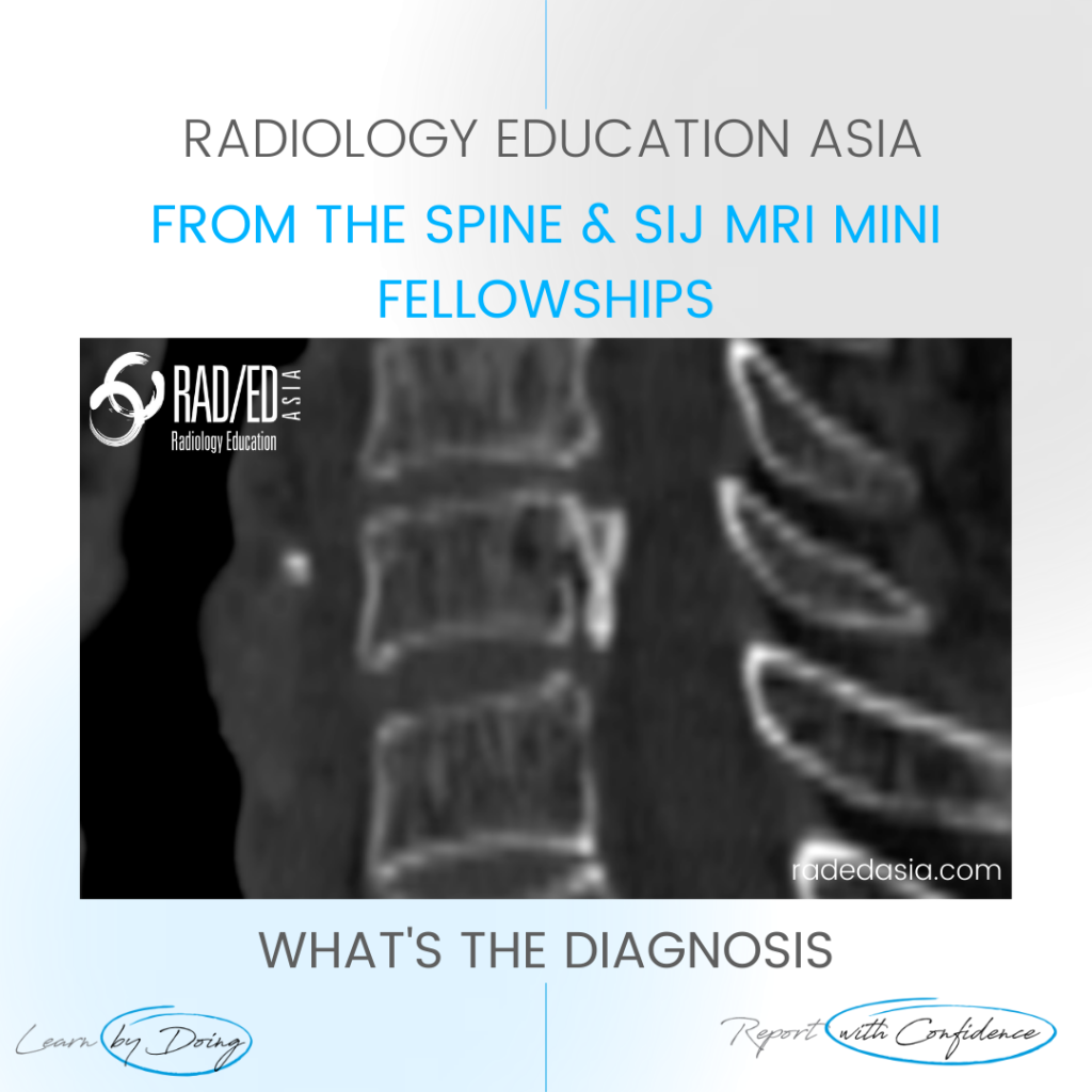
OPLL SPINE OSSIFICATION POSTERIOR LONGITUDINAL LIGAMENT CT DISCUSSION ON OPLL SPINE CT There is localised, linear ossification present at the posterior margin of the C6 vertebra.   This has a typical appearance of OPLL (Ossification of the Posterior Longitudinal Ligament).   The cervical spine is the most common location.   It is important to follow …

OPLL SPINE OSSIFICATION POSTERIOR LONGITUDINAL LIGAMENT CT (VIDEO) Read More »

ischiofemoral impingement radiology mri hip

ISCHIOFEMORAL IMPINGEMENT RADIOLOGY HIP MRI (VIDEO)

ISCHIOFEMORAL IMPINGEMENT HIP MRI RADIOLOGY ISCHIOFEMORAL IMPINGEMENT: WHY DOES IT OCCUR Ischiofemoral impingement is a result of the quadratus femoris muscle being compressed between the ischial tuberosity and the posterior femur resulting initially in quadratus femoris oedema but can progress to tears and severe atrophy. ISCHIOFEMORAL IMPINGEMENT: WHAT ARE THE FINDINGS Initially there is oedema …

ISCHIOFEMORAL IMPINGEMENT RADIOLOGY HIP MRI (VIDEO) Read More »

learn mri knee mucoid degen radedasia

ACL MUCOID DEGENERATION RADIOLOGY MRI

ACL MUCOID DEGENERATION RADIOLOGY MRI DISCUSSION Mucoid Degeneration. These findings indicate Mucoid Degeneration of the ACL rather than a tear.   This is a characteristic appearance of ACL mucoid degeneration where the ACL is expanded and hyperintense but is still continuous with normal alignment. . WHAT ARE THE FINDINGS The ACL is enlarged and hyperintense …

ACL MUCOID DEGENERATION RADIOLOGY MRI Read More »

retroachilles bursa bursitis mri radiology ankle

RETROACHILLES BURSA BURSITIS MRI RADIOLOGY ANKLE

RETROACHILLES BURSA BURSITIS MRI ANKLE DISCUSSION ABOUT THE RETROACHILLES BURSA The retroachilles bursa lies between skin and Achilles tendon.   It’s not usually seen as its an adventitial bursa caused by chronic friction.   It is Uncommonly enlarged. WHERE TO LOOK FOR THE RETROACHILLES BURSA Best seen on axial and sagittal scans.   Look posterior …

RETROACHILLES BURSA BURSITIS MRI RADIOLOGY ANKLE Read More »

ankle mri peroneus brevis tendon tear split

PERONEUS BREVIS TENDON TEAR MRI ANKLE MRI

PERONEUS BREVIS TENDON TEAR MRI DISCUSSION: WHAT'S THE dX Peroneus Brevis Tendon Tear: MRI demonstrates a peroneus brevis tendon tear.  There is a longitudinal split resulting in two components of the Peroneus Brevis tendon (Pink arrows). Peroneus Longus (Blue arrow) is normal. WHAT ARE THE FINDINGS There should only be two tendons in the peroneal …

PERONEUS BREVIS TENDON TEAR MRI ANKLE MRI Read More »

shoulder synovitis mri rheumatoid arthritis subacromaial bursa radedasia

SHOULDER SYNOVITIS MRI RHEUMATOID ARTHRITIS

SHOULDER SYNOVITIS MRI RHEUMATOID ARTHRITIS DISCUSSION: WHAT'S THE Dx? Extensive synovitis and ACJ erosions from Rheumatoid arthritis. Having synovitis in multiple locations (even if there are no erosions) should raise the possibility of an inflammatory abnormality.   Thank you to Dr Joe Thomas (Rheumatologist) for the case. WHAT ARE THE FINDINGS? Image 1: The first …

SHOULDER SYNOVITIS MRI RHEUMATOID ARTHRITIS Read More »

mri acl tear pivot shift injury bone contusion pattern osteochondral fracture knee

ACL TEAR PIVOT SHIFT INJURY BONE CONTUSION PATTERN OSTEOCHONDRAL FRACTURE MRI KNEE

ACL TEAR PIVOT SHIFT INJURY BONE CONTUSION PATTERN OSTEOCHONDRAL FRACTURE MRI WHAT'S THE Dx? Pivot shift bone bruising of the lateral femoral condyle and an osteochondral depression fracture secondary to an ACL tear.   Characteristic bone contusion pattern seen with complete ACL tear.   The osteochondral fracture is seen with the flattening of the cortical …

ACL TEAR PIVOT SHIFT INJURY BONE CONTUSION PATTERN OSTEOCHONDRAL FRACTURE MRI KNEE Read More »

spine trauma imaging facet fracture dislocation radiology ct

FACET FRACTURE SPINE RADIOLOGY CT TRAUMA IMAGING

FACET FRACTURE RADIOLOGY CT DISCUSSION FACET FRACTURE CT APPEARANCE The facet is fractured (Pink arrows) but there is no evidence on this image of facet subluxation or dislocation.   However the facet joint is widened inferiorly (Green arrow) indicating that the facet capsule must be torn. FACET FRACTURE RADIOLOGY CT: SLIDER TO VIEW IMAGES ARE …

FACET FRACTURE SPINE RADIOLOGY CT TRAUMA IMAGING Read More »

This site is intended for Medical Professionals only. Use of this site is governed by our Terms of Service and Privacy Statement which can be found by clicking on the links. Please accept before proceeding to the website.
error: Right Click not available.