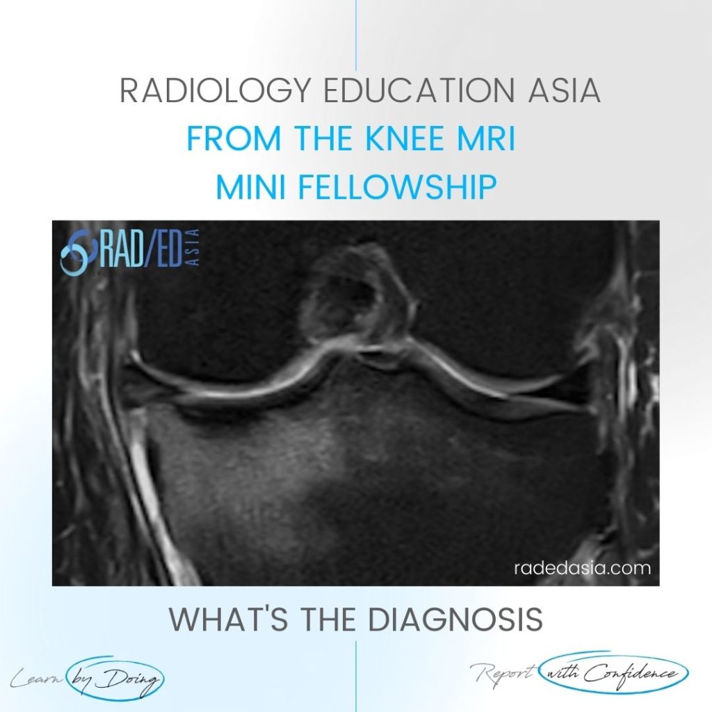
- Subchondral or subcortical fractures of the knee can occur in the femoral condyles or tibial plateau.
- The key to the diagnosis is seeing a linear low signal line adjacent to and paralleling the cortex without any cortical defect or break in the acute phase.
- There is usually significant bone marrow oedema spreading around the fracture line.
- An older term for this appearance is SONK (Spontaneous Osteonecrosis of the Knee) however the pathology is now recognised to commence with a subcortical fracture.
- More chronically osteonecrosis can occur in the region of the fracture with collapse of the cortex.
- When this occurs there will be contour deformity of the cortex.

- Extensive bone marrow oedema tibial plateau (Yellow arrows) and,
- Linear low signal subcortical line which is low signal on T2FS and T1 (Green arrows) which is a subcortical fracture.
- Additional and not directly related extrusion of the meniscus (Pink arrow).

Learn more about KNEE Imaging in our ONLINE or ONSITE
Guided MRI KNEE Mini-Fellowship.
More by clicking on the images below.
#radiology #radiologist #radiologia #mri #kneemri #mskmri #msk #mskrad #mskradiology #imaging #frcr #sportsmed #radiologyresident #foamrad #emergencydepartment #ortho #ct #radiologystudent #trauma #radedasia #radiologycme #radiologyeducation #radiologycases #rheumatology #arthritis #painphysician #chiropractic #physiotherap #kneesurgeon
#radedasia #mri #mskmri #radiology


