SPINE

SPINE MRI Learn Onlinelearn spine mri workshop online

All our posts on Spine MRI Radiology . We regularly post on Spine MRI with all the posts on this page.

Spine MRI Radiology can be difficult due to the complex anatomy and the many pathologies that occur. In our posts we have concise explanation of the Spine MRI anatomy, the macroscopic pathology that’s relevant to the Imaging and the Imaging findings in various pathologies.

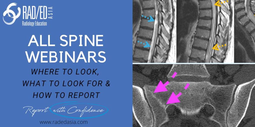
Learn where to Look, What to Look for and How to Report More Confidently

learn spine imaging mri ct xray radiology conference course trauma anatomy degeneration arthritis

SPINE IMAGING MRI CT XRAY RADIOLOGY CONFERENCE COURSE LEARN

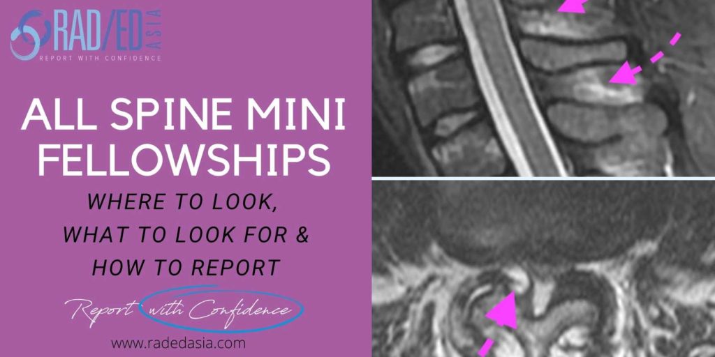
Home SPINE IMAGING COURSE: SPINE MRI CT XRAY RADIOLOGY Spine Imaging Courses. Learn about imaging the spine in our Guided Spine Imaging Mini Fellowships.  30 Day courses that cover the Anatomy, Pathology that assists in the diagnosis and Imaging of spinal abnormalities. Where to Look, What to Look for and How to Report…With Confidence. OUR …

SPINE IMAGING MRI CT XRAY RADIOLOGY CONFERENCE COURSE LEARN Read More »

spine trauma imaging facet fracture dislocation radiology ct

FACET FRACTURE SPINE RADIOLOGY CT TRAUMA IMAGING

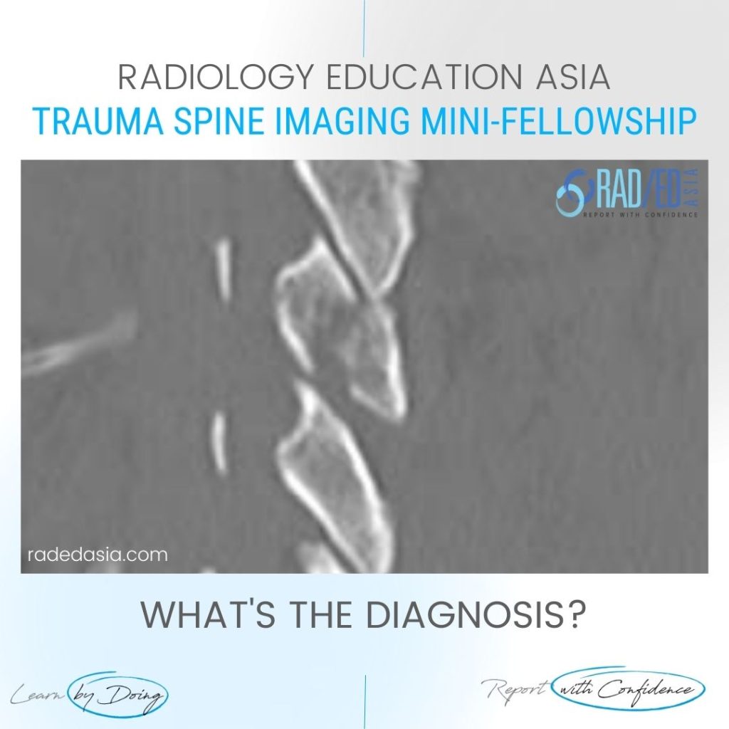
FACET FRACTURE RADIOLOGY CT DISCUSSION FACET FRACTURE CT APPEARANCE The facet is fractured (Pink arrows) but there is no evidence on this image of facet subluxation or dislocation.   However the facet joint is widened inferiorly (Green arrow) indicating that the facet capsule must be torn. FACET FRACTURE RADIOLOGY CT: SLIDER TO VIEW IMAGES ARE …

FACET FRACTURE SPINE RADIOLOGY CT TRAUMA IMAGING Read More »

spine ligaments mri injury radiology interspinous trauma

SPINE LIGAMENTS MRI INJURY RADIOLOGY TRAUMA INTERSPINOUS

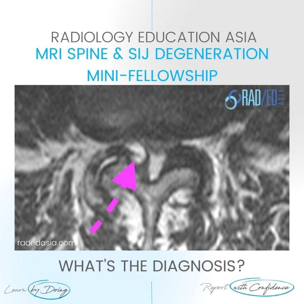
SPINE LIGAMENTS MRI INJURY RADIOLOGY TRAUMA INTERSPINOUS WHAT'S THE Dx: INTERSPINOUS LIGAMENT TRAUMA The MRI demonstrates trauma to the interspinous ligament with increased T2FS signal in the region of the interspinous ligament.   The interspinous ligament lies between the spinous processes.   Oedema indicates trauma to the region but is also a clue to the …

SPINE LIGAMENTS MRI INJURY RADIOLOGY TRAUMA INTERSPINOUS Read More »

vertebral artery dissection radiology mri diagnosis

VERTEBRAL ARTERY DISSECTION RADIOLOGY MRI DIAGNOSIS

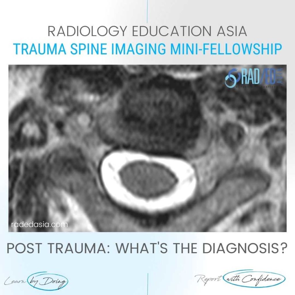
VERTEBRAL ARTERY DISSECTION RADIOLOGY MRI DIAGNOSIS MRI VERTEBRAL ARTERY OCCULSION: DISCUSSION VERTEBRAL ARTERY OCCULSION   With spinal trauma its essential to look at the signal of the vertebral arteries as there can be dissection.   In most cases we don’t do a routine MRA so being able to spot a dissection on the standard scans …

VERTEBRAL ARTERY DISSECTION RADIOLOGY MRI DIAGNOSIS Read More »

mri spine rheumatoid arthritis basilar invagination radiology education asia

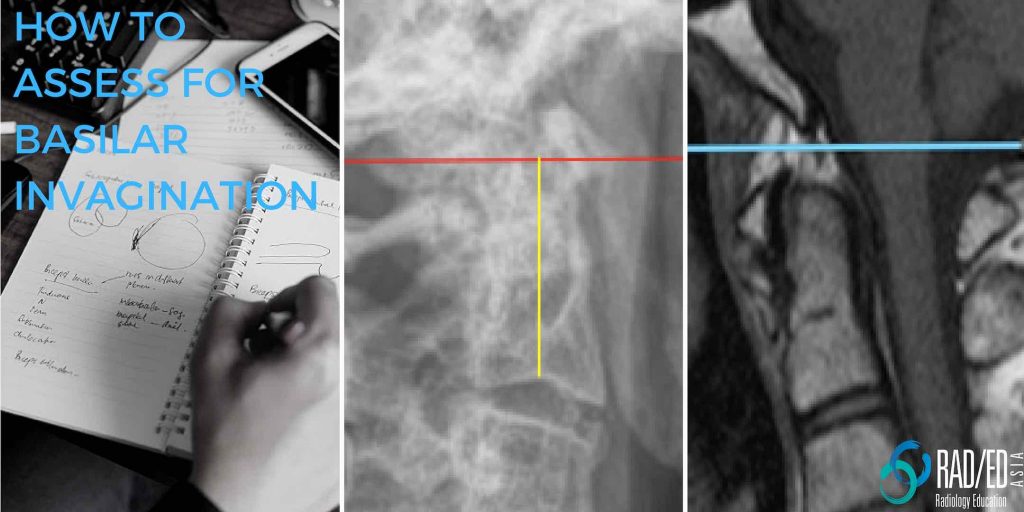
BASILAR INVAGINATION RADIOLOGY RHEUMATOID ARTHRITIS FIVE WAYS TO ASSESS

BASILAR INVAGINATION MRI RHEUMATOID ARTHRITIS WHAT IS BASILAR INVAGINATION / IMPRESSION BASILAR INVAGINATION RADIOLOGY RHEUMATOID ARTHRITIS: Subluxation at C1/2 in Rheumatoid Arthritis can occur in three directions. Axial (Horizontal). Vertical and, Lateral. Vertical subluxation is when C2 moves proximally towards the foramen magnum and is called Basilar Invagination or Basilar Impression. Basilar Invagination is when …

BASILAR INVAGINATION RADIOLOGY RHEUMATOID ARTHRITIS FIVE WAYS TO ASSESS Read More »

spine mri ankylosing spondylitis spondyloarthritis SpA rheumatologist rheumatology radiology

MRI SPINE ANKYLOSING SPONDYLITIS SpA SPONDYLOARTHRITIS FOR RHEUMATOLOGISTS: HOW TO ASSESS: PART 2 WHAT DOES THE SIGNAL MEAN

MRI SPINE FOR RHEUMATOLOGISTS & (RADIOLOGISTS) In this video below we look at how to assess a MRI Spine Ankylosing Spondylitis & Spondyloarthritis (SpA ) for Rheumatologists (and Radiologists ) and look at the SIGNAL CHANGES YOU NEED TO KNOW AND WHAT  THEY MEAN. This is the 2nd part of a 3 part series on …

MRI SPINE ANKYLOSING SPONDYLITIS SpA SPONDYLOARTHRITIS FOR RHEUMATOLOGISTS: HOW TO ASSESS: PART 2 WHAT DOES THE SIGNAL MEAN Read More »

MRI Spine Rheumatologist rheumatology Ankylosing Spondylitis Spondyloarthropathy radedasia

MRI SPINE RHEUMATOLOGY FOR RHEUMATOLOGISTS: HOW TO ASSESS ANKYLOSING SPONDYLITIS AND SPONDYLOARTHROPATHY PART 1

MRI SPINE FOR RHEUMATOLOGISTS & (RADIOLOGISTS) How to assess Ankylosing Spondylitis and Spondyloarthropathy Part 1: The Sequences you need to know. Rheumatologists should be able to assess a MRI Spine for changes of Ankylosing Spondylitis and Spondyloarthropathy. We have a 3 part series for Rheumatologists (and Radiologists 😀) on how to have a structure to assess …

MRI SPINE RHEUMATOLOGY FOR RHEUMATOLOGISTS: HOW TO ASSESS ANKYLOSING SPONDYLITIS AND SPONDYLOARTHROPATHY PART 1 Read More »

cord signal change spondylotic myelopathy mri

CORD SIGNAL CHANGE MRI SPONDYLOTIC MYELOPATHY

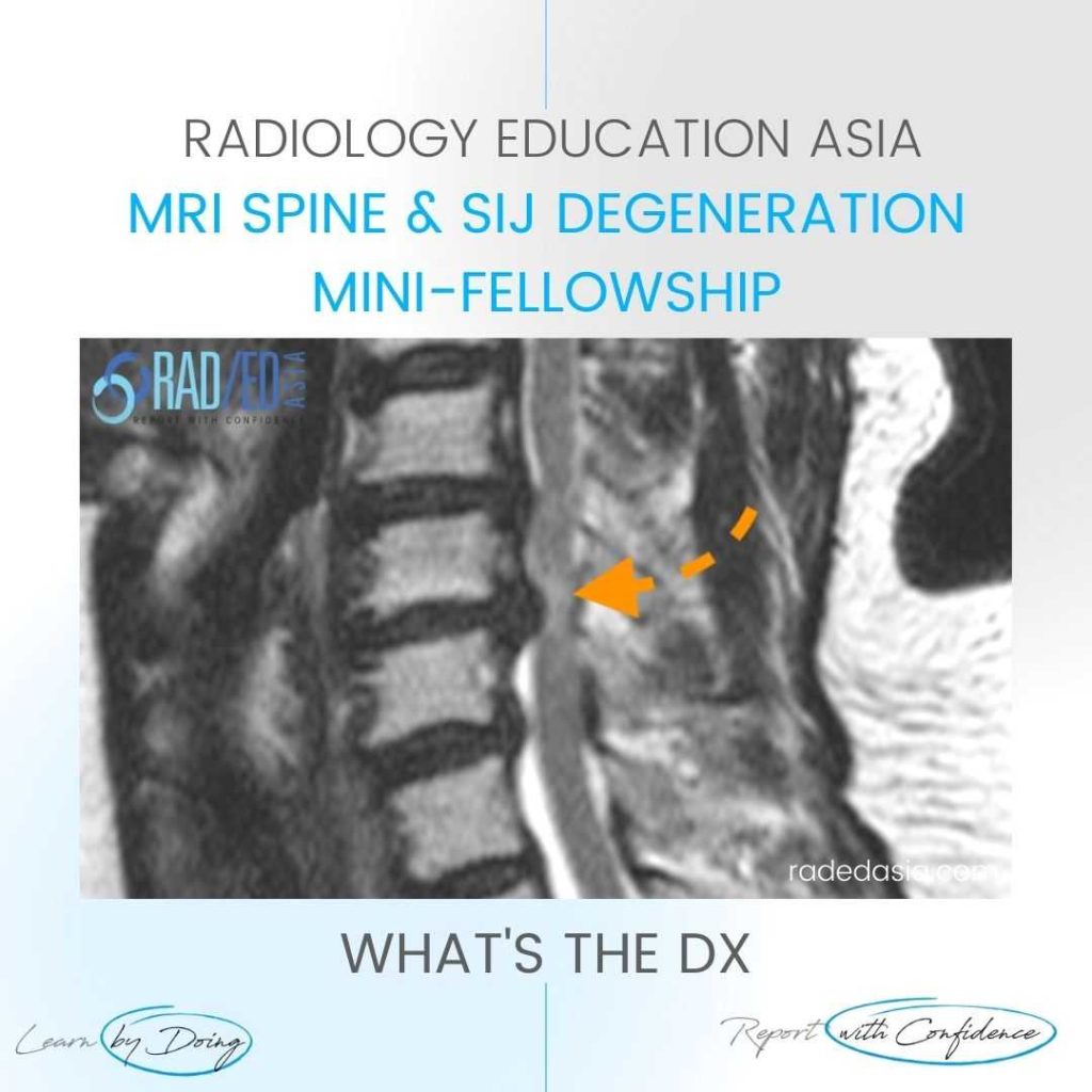
SPONDYLOTIC MYELOPATHY MRI RADIOLOGY DISCUSSION SPONDYLOTIC MYELOPATHY   Spondylotic Myelopathy is a term used when there is increased cord signal due to extrinsic compression.   Patients will often be symptomatic with cord related symptoms.   By using the term you are indicating that the patient’s symptoms can potentially be reversed by relieving the compression. WHAT …

CORD SIGNAL CHANGE MRI SPONDYLOTIC MYELOPATHY Read More »

This site is intended for Medical Professionals only. Use of this site is governed by our Terms of Service and Privacy Statement which can be found by clicking on the links. Please accept before proceeding to the website.
error: Right Click not available.