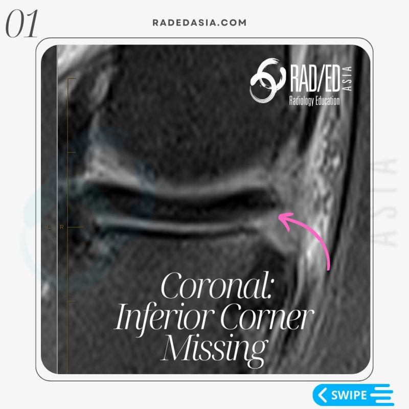
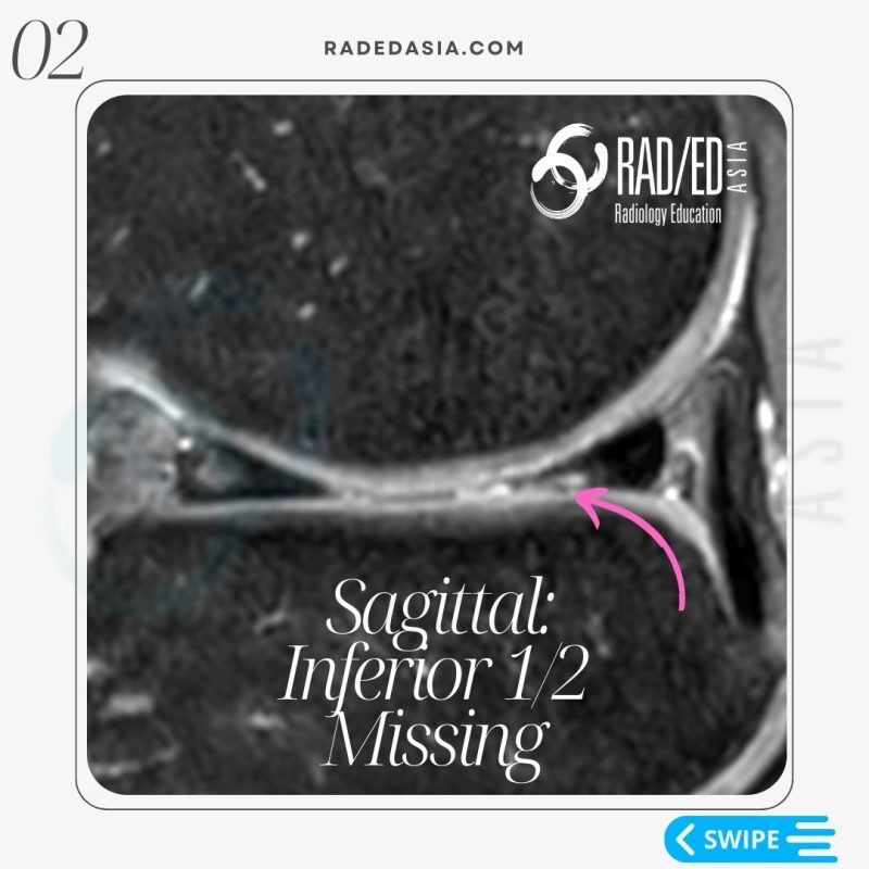
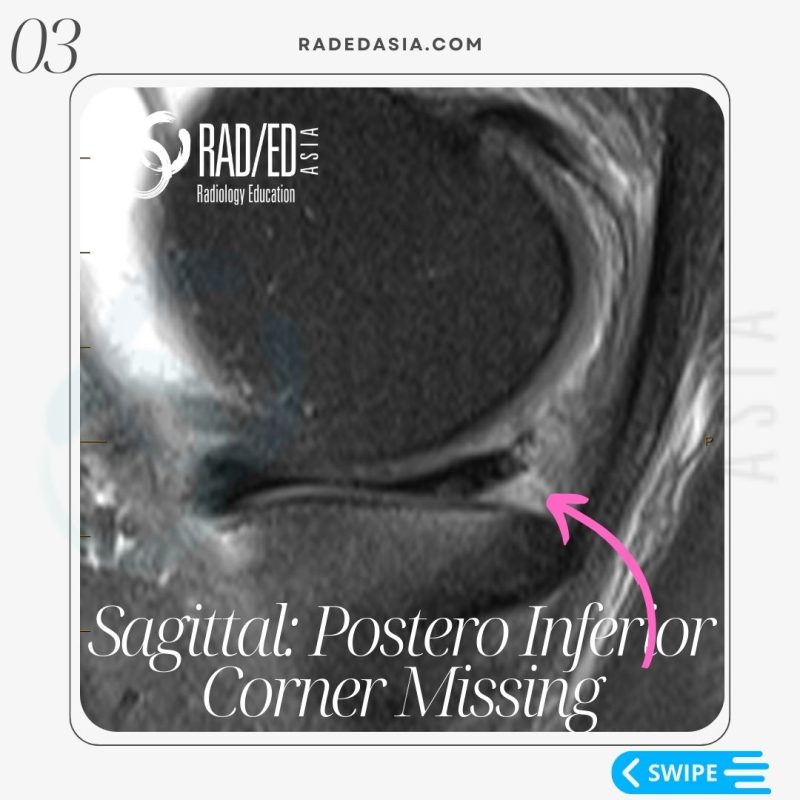
• In a Normal meniscus the superior and inferior halves should be symmetric.
• In all three cases there is localised loss of " volume" of the meniscus.
• This gives the meniscus an asymmetric look.
• Asymmetry in a meniscus is always abnormal and we need to look further to work out why..

- Meniscal tears are not just linear.
- Absence of a portion of a meniscus is also a tear.
- Know the normal appearance of different parts of the meniscus.
- Then look harder at any areas of asymmetry.

Learn more about KNEE Imaging in our ONLINE or ONSITE
Guided MRI KNEE Mini-Fellowship.
More by clicking on the images below.
- Join our WhatsApp RadEdAsia community for regular educational posts at this link: https://bit.ly/radedasiacommunity
- Get our weekly email with all our educational posts: https://bit.ly/whathappendthisweek
#radedasia #mri #mskmri #radiology


