SEO

mri musculotendinous junction cysts and delaminating rotator cuff tears

MRI DELAMINATION ROTATOR CUFF TEARS MUSCULOTENDINOUS JUNCTION CYSTS

MRI DELAMINATING ROTATOR CUFF TEARS & MUSCULOTENDINOUS JUNCTION CYSTS On MRI Rotator cuff delaminating tears can result in Musculotendinous Junction Cysts.   These Musculotendinous junction cysts are seen in the rotator cuff tendons, mostly in Supraspinatus and less commonly in infraspinatus.   In this post we look at the appearance of both Delaminating Rotator cuff …

MRI DELAMINATION ROTATOR CUFF TEARS MUSCULOTENDINOUS JUNCTION CYSTS Leer más »

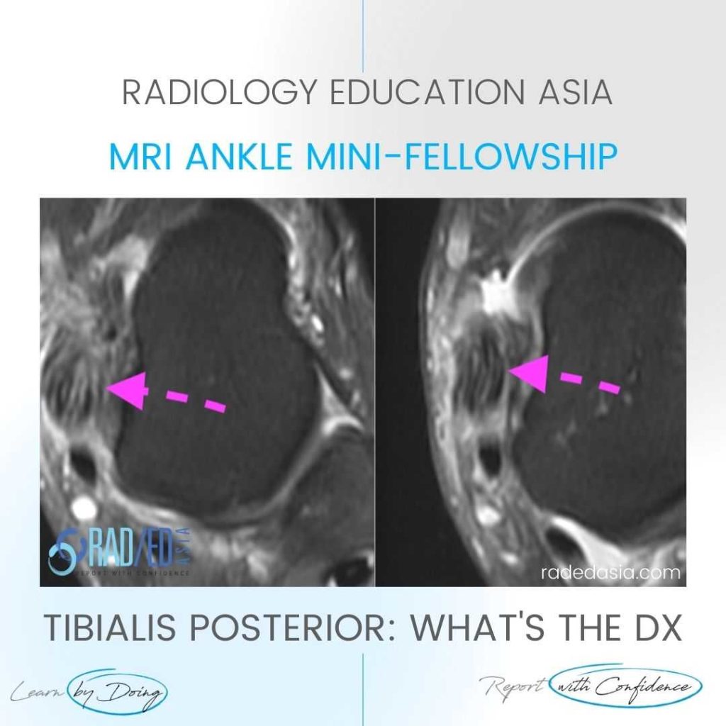
tibialis posterior tendinitis tendinopathy tear mri radiology

TIBIALIS POSTERIOR TENDINITIS TENDINOPATHY TEARS RADIOLOGY MRI (VIDEO)

TIBIALIS POSTERIOR TENDINITIS TENDINOPATHY TEARS RADIOLOGY MRI WHAT ARE THE FINDINGS The tibialis posterior tendon is enlarged and increased in signal. DIAGNOSIS & DISCUSSION TIBIALIS POSTERIOR TENDINOPATHY AND PARTIAL TEAR. Tibialis posterior tendinopathy and delamination / partial tears. The normal tibialis posterior tendon should be uniformly low signal. Enlargement and or increased intermediate signal indicates …

TIBIALIS POSTERIOR TENDINITIS TENDINOPATHY TEARS RADIOLOGY MRI (VIDEO) Leer más »

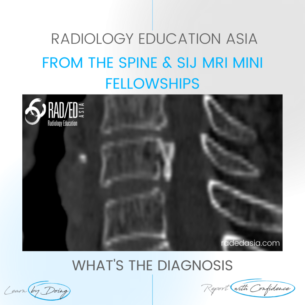
opll spine ossification posterior longitudinal ligament ct mri xray radiology (1)

OPLL SPINE OSSIFICATION POSTERIOR LONGITUDINAL LIGAMENT CT (VIDEO)

OPLL SPINE OSSIFICATION POSTERIOR LONGITUDINAL LIGAMENT CT DISCUSSION ON OPLL SPINE CT There is localised, linear ossification present at the posterior margin of the C6 vertebra.   This has a typical appearance of OPLL (Ossification of the Posterior Longitudinal Ligament).   The cervical spine is the most common location.   It is important to follow …

OPLL SPINE OSSIFICATION POSTERIOR LONGITUDINAL LIGAMENT CT (VIDEO) Leer más »

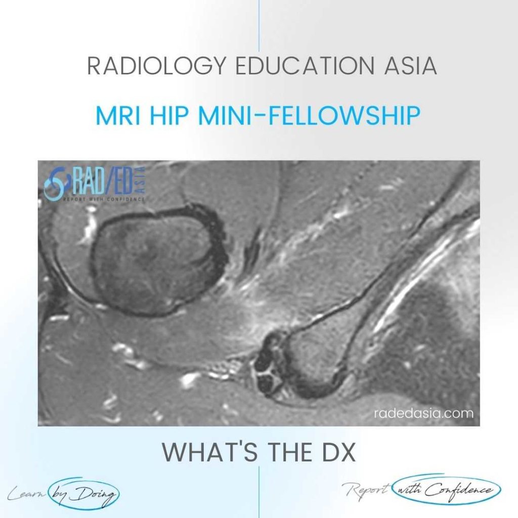
ischiofemoral impingement radiology mri hip

ISCHIOFEMORAL IMPINGEMENT RADIOLOGY HIP MRI (VIDEO)

ISCHIOFEMORAL IMPINGEMENT HIP MRI RADIOLOGY ISCHIOFEMORAL IMPINGEMENT: WHY DOES IT OCCUR Ischiofemoral impingement is a result of the quadratus femoris muscle being compressed between the ischial tuberosity and the posterior femur resulting initially in quadratus femoris oedema but can progress to tears and severe atrophy. ISCHIOFEMORAL IMPINGEMENT: WHAT ARE THE FINDINGS Initially there is oedema …

ISCHIOFEMORAL IMPINGEMENT RADIOLOGY HIP MRI (VIDEO) Leer más »

osteitis condensans illii mri spine radiology

OSTEITIS CONDENSANS ILII MRI SPINE RADIOLOGY (VIDEO)

OSTEITIS CONDENSANS ILII MRI DIAGNOSIS OSTEITIS CONDENSANS ILII DISCUSSION OSTEITIS CONDENSANS ILII a common finding that may be confusing to diagnose on MRI.   Look for low signal on all sequences   The findings are predominantly of the iliac side of the SIJ but may be seen additionally on the sacral side.   It doesn’t occur …

OSTEITIS CONDENSANS ILII MRI SPINE RADIOLOGY (VIDEO) Leer más »

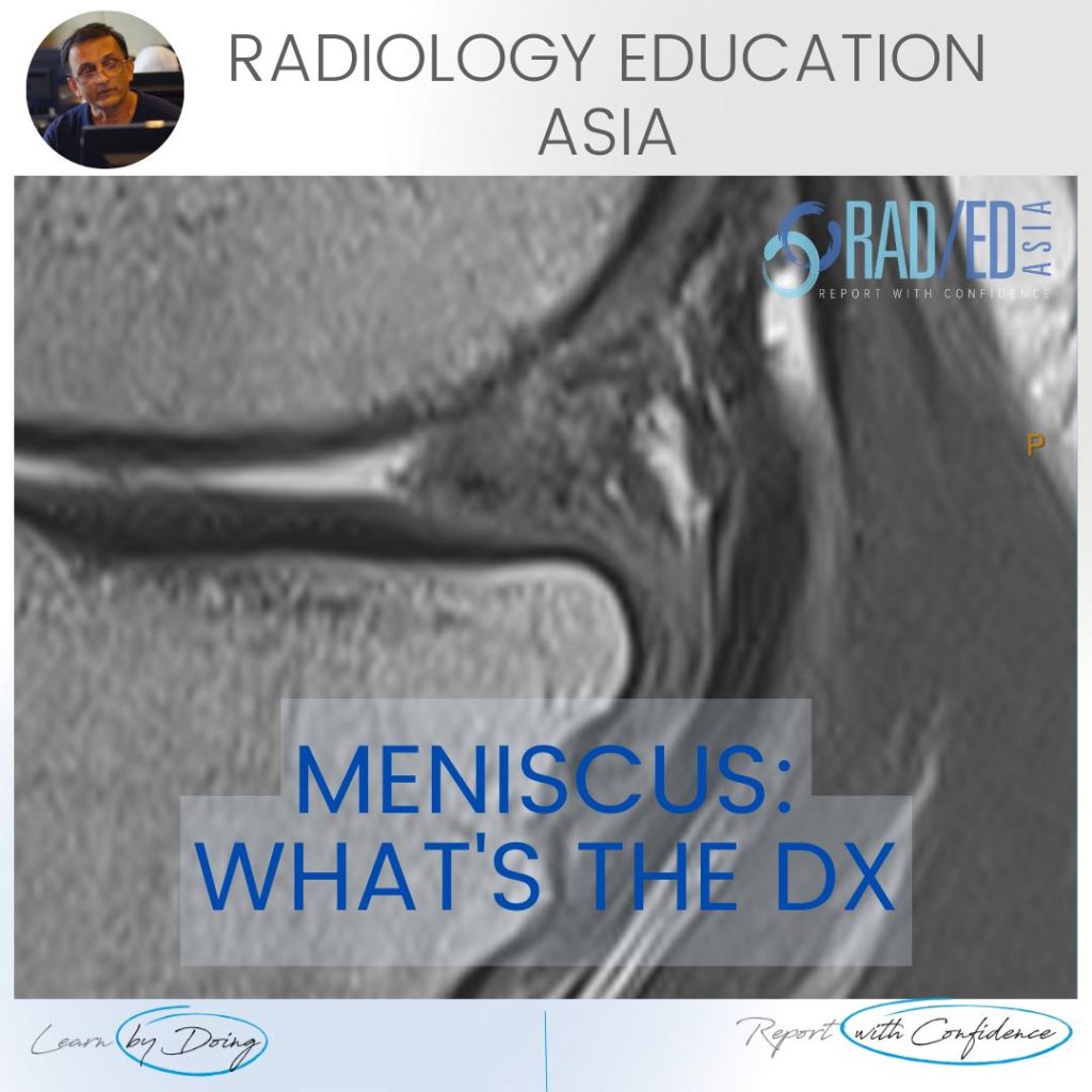
meniscus tear maceration degeneration mri knee radedasia

MENISCUS TEAR MACERATION DEGENERATION MRI KNEE RADIOLOGY (VIDEO)

MENISCUS MACERATION DEGENERATION MRI KNEE WHAT ARE THE FINDINGS MENISCUS MACERATION DEGENERATION MRI KNEE   Generalized increase in meniscal signal. Intermediate increase in signal on PD and PD/T2FS. The meniscus generally retains it’s shape but is ill defined. Due to the degeneration its more likely to tear and it also stops functioning like a normal …

MENISCUS TEAR MACERATION DEGENERATION MRI KNEE RADIOLOGY (VIDEO) Leer más »

annular fissure tear mri radiology lumbar spine radedasia

ANNULAR FISSURE TEAR MRI RADIOLOGY LUMBAR SPINE (VIDEO)

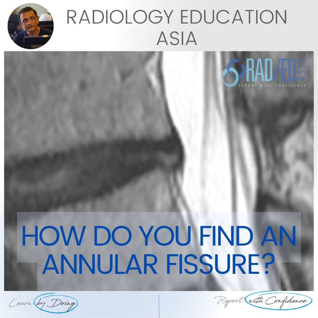
ANNULAR FISSURE TEAR MRI RADIOLOGY HOW DO YOU FIND AN ANNULAR FISSURE ANNULAR FISSURE ON MRI Annular fissures on MRI are relatively common. Whilst they don’t result in radiculopathy, they can be a cause of back pain and are important to diagnose and report.   What does an annular fissure look like and where to …

ANNULAR FISSURE TEAR MRI RADIOLOGY LUMBAR SPINE (VIDEO) Leer más »

stress fracture fatigue insufficiency fracture radiology mri radedasia

STRESS FRACTURE FATIGUE FRACTURE AND INSUFFICIENCY FRACTURE WHAT IS THE DIFFERENCE (VIDEO)

FATIGUE FRACTURE AND INSUFFICIENCY FRACTURE: WHAT'S THE DIFFERENCE DISCUSSION The difference lies in the underlying bone metabolism. Is it normal or not. A Fatigue fracture is  stress type fracture due to repetitive overload, with normal bone metabolism (eg a March Fracture of the metatarsal in new army recruits). An Insufficiency fracture is a stress type …

STRESS FRACTURE FATIGUE FRACTURE AND INSUFFICIENCY FRACTURE WHAT IS THE DIFFERENCE (VIDEO) Leer más »

ankle impingement radiology anterior xray mri radedasia

ANKLE IMPINGEMENT RADIOLOGY ANTERIOR X-RAY (VIDEO)

ANKLE IMPINGEMENT RADIOLOGY ANTERIOR X-RAY DISCUSSION Ankle impingement can occur at multiple sites. One of the more common ones is Anterior.   The presence of Ankle impingement is ultimately a clinical diagnosis which is based on the patient’s symptoms and imaging findings, not just the imaging findings.   So in the report we describe the …

ANKLE IMPINGEMENT RADIOLOGY ANTERIOR X-RAY (VIDEO) Leer más »

Este sitio está destinado únicamente a profesionales médicos. El uso de este sitio se rige por nuestra Términos de servicio y Declaracion de privacidad que se puede encontrar haciendo clic en los enlaces. Acepte antes de continuar con el sitio web.
error: Clic derecho no disponible.