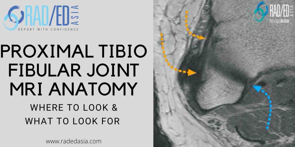
MRI KNEE ANATOMY: PROXIMAL TIBIOFIBULAR JOINT
PROXIMAL TIBIOFIBULAR JOINT MRI ANATOMY MRI PROXIMAL TIBIO FIBULAR JOINT ANATOMY The proximal tibiofibular joint is often overlooked when assessing a knee MRI. This is the first in a series on MRI of the proximal tibiofibular joint (PTFJ) beginning with the anatomy of the ligaments that stabilize the PTFJ. WHAT'S THE PROBLEM Often overlooked when …