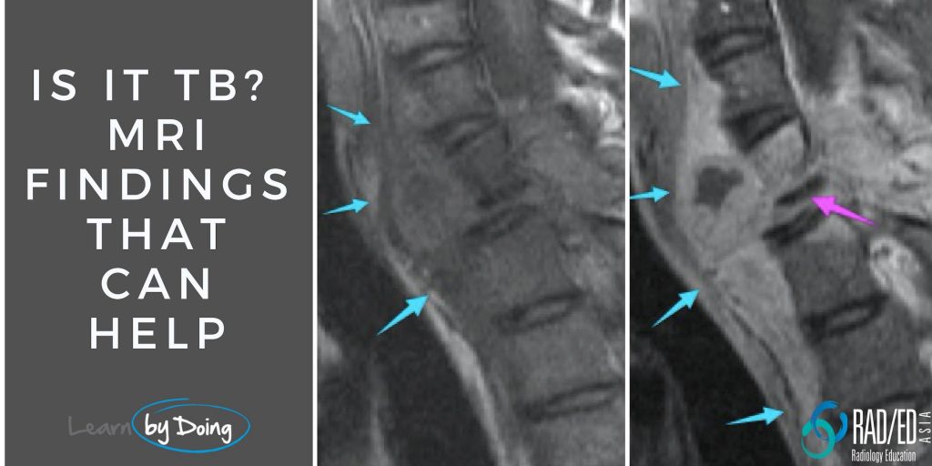
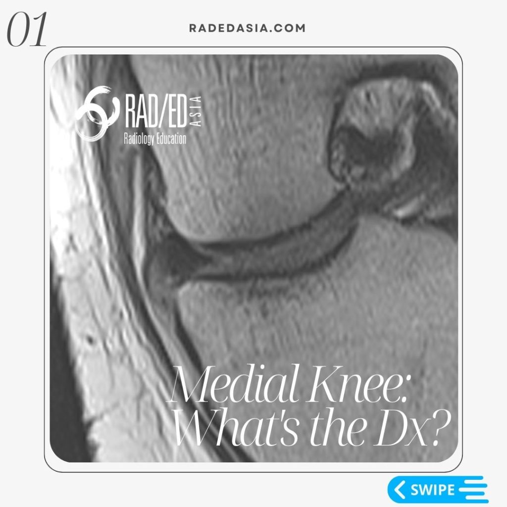
Dr. Ravi Padmanabhan

Coracoid Process tendon attachment coracobrachialis short head biceps

CORACOID PROCESS TENDON ATTACHMENT MRI CORACOBRACHIALIS & SHORT HEAD OF BICEPS (VIDEO)

CORACOID PROCESS TENDON ATTACHMENT MRI CORACOBRACHIALIS & SHORT HEAD OF BICEPS HOW TO FIND CORACOBRACHIALIS & SHORT HEAD OF BICEPS:   There are 3 tendons that attach to the coracoid process. Pectoralis Minor. Coracobrachialis and, The short Head of Biceps.   In this video, we look at how to identify on MRI the Coracobrachialis and …

CORACOID PROCESS TENDON ATTACHMENT MRI CORACOBRACHIALIS & SHORT HEAD OF BICEPS (VIDEO) Leer más »

coracoid process tendon attachment pectoralis minor mri shoulder radedasia

CORACOID PROCESS TENDON ATTACHMENT PECTORALIS MINOR MRI (VIDEO)

CORACOID PROCESS TENDON ATTACHMENT PECTORALIS MINOR MRI HOW TO FIND PECTORALIS MINOR:   There are 3 tendons that attach to the coracoid process. Pectoralis Minor. Coracobrachialis and, The short Head of Biceps.   In this video we start by looking at how to identify on MRI the Pectoralis Minor muscle and tendon on a Shoulder …

CORACOID PROCESS TENDON ATTACHMENT PECTORALIS MINOR MRI (VIDEO) Leer más »

tb spine tuberculosis mri radiology education asia

MRI TB SPINE RADIOLOGY TUBERCULOSIS: WHAT DOES SPINAL TB LOOK LIKE ON MRI

MRI TB SPINE RADIOLOGY TUBERCULOSIS MRI TB SPINE RADIOLOGY TUBERCULOSIS: WHAT DOES SPINAL TB LOOK LIKE ON MRI When there is spinal infection, a common question from the referrer will be ” Is it TB? “.  It can be difficult to tell as MRI findings in spinal TB can look exactly like bacterial spondylodiscitis but …

MRI TB SPINE RADIOLOGY TUBERCULOSIS: WHAT DOES SPINAL TB LOOK LIKE ON MRI Leer más »

ankle mri calcaneofibular ligament tear radedasia

CALCANEOFIBULAR LIGAMENT TEAR MRI ANKLE

CALCANEOFIBULAR LIGAMENT TEAR MRI ANKLE CALCANEOFIBULAR LIGAMENT NORMAL MRI The Calcaneofibular ligament is a small lateral ankle ligament that is often torn.   Its normal MRI appearance is a thin low signal structure (Pink arrows) that extends from the calcaneum to the fibula and passes deep to the peroneal tendons (Green arrows).   CALCANEOFIBULAR LIGAMENT …

CALCANEOFIBULAR LIGAMENT TEAR MRI ANKLE Leer más »

knee infrapatellar plica syndrome radiology mri radedasia

KNEE INFRAPATELLAR PLICA SYNDROME RADIOLOGY MRI (VIDEO)

KNEE INFRAPATELLAR PLICA SYNDROME RADIOLOGY MRI DISCUSSION ON KNEE INFRAPATELLAR PLICA PLICAS IN THE KNEE The infrapatellar plica is one of 4 plicas in the knee: Medial Patella Plica. Suprapatellar Plica. Infrapatellar plica. Lateral Patellar plica. INFRAPATELLAR PLICA ANATOMY Attaches to anterior intercondylar notch and inferior pole patella.   Runs through Hoffa’s Fat Pad.   …

KNEE INFRAPATELLAR PLICA SYNDROME RADIOLOGY MRI (VIDEO) Leer más »

bony bankart xray ct radiology glenoid fracture shoulder dislocation

SHOULDER DISLOCATION BONY BANKART GLENOID FRACTURE XRAY CT (VIDEO)

BONY BANKART LESION RADIOLOGY XRAY FINDING 1: BONE FRAGMENT Image Above: There is a thin fragment of bone at the inferior margin of the glenoid. FINDING 2: GLENOID FLATTENING Image Above: The inferior margin of the glenoid should normally be rounded and ovoid. Here it is flattened which is abnormal. DISCUSSION Bony Bankart lesions on …

SHOULDER DISLOCATION BONY BANKART GLENOID FRACTURE XRAY CT (VIDEO) Leer más »

superficial siderosis mri arachnoiditis arachmoid cyst radedasia

SUPERFICIAL SIDEROSIS MRI CORD AND SPINE: ARACHNOIDITIS & ARACHNOID CYST (VIDEO)

SUPERFICIAL SIDEROSIS: ARACHNOIDITIS & CYST Superficial Siderosis MRI Cord and Spine: Complications and Associations. Superficial Siderosis can affect the cord, nerve roots and the CSF space in the thecal sac. Here is what to look for and where to look to not miss this diagnosis. SUPERFICIAL SIDEROSIS MRI COMPLICATIONS TO LOOK FOR 1. ARACHNOIDITIS: WHY …

SUPERFICIAL SIDEROSIS MRI CORD AND SPINE: ARACHNOIDITIS & ARACHNOID CYST (VIDEO) Leer más »

osgood schlatter xray radiology radedasia

OSGOOD SCHLATTER RADIOLOGY X-RAY (VIDEO)

OSGOOD SCHLATTER RADIOLOGY X-RAY FINDINGS There are three findings in the x-ray that make the diagnosis ofOsgood Schlatter. Bone avulsion from tibial tuberosity.  Soft tissue swelling over tibial tuberosity.  Increased density deep infra patella bursa from fluid/inflammation. OSGOOD SCHLATTER X-RAY FINDINGS: VIEW VIDEO https://youtu.be/qG4b7ik9G3I If your Browser is blocking the video, Please view it on …

OSGOOD SCHLATTER RADIOLOGY X-RAY (VIDEO) Leer más »

Este sitio está destinado únicamente a profesionales médicos. El uso de este sitio se rige por nuestra Términos de servicio y Declaracion de privacidad que se puede encontrar haciendo clic en los enlaces. Acepte antes de continuar con el sitio web.
error: Clic derecho no disponible.