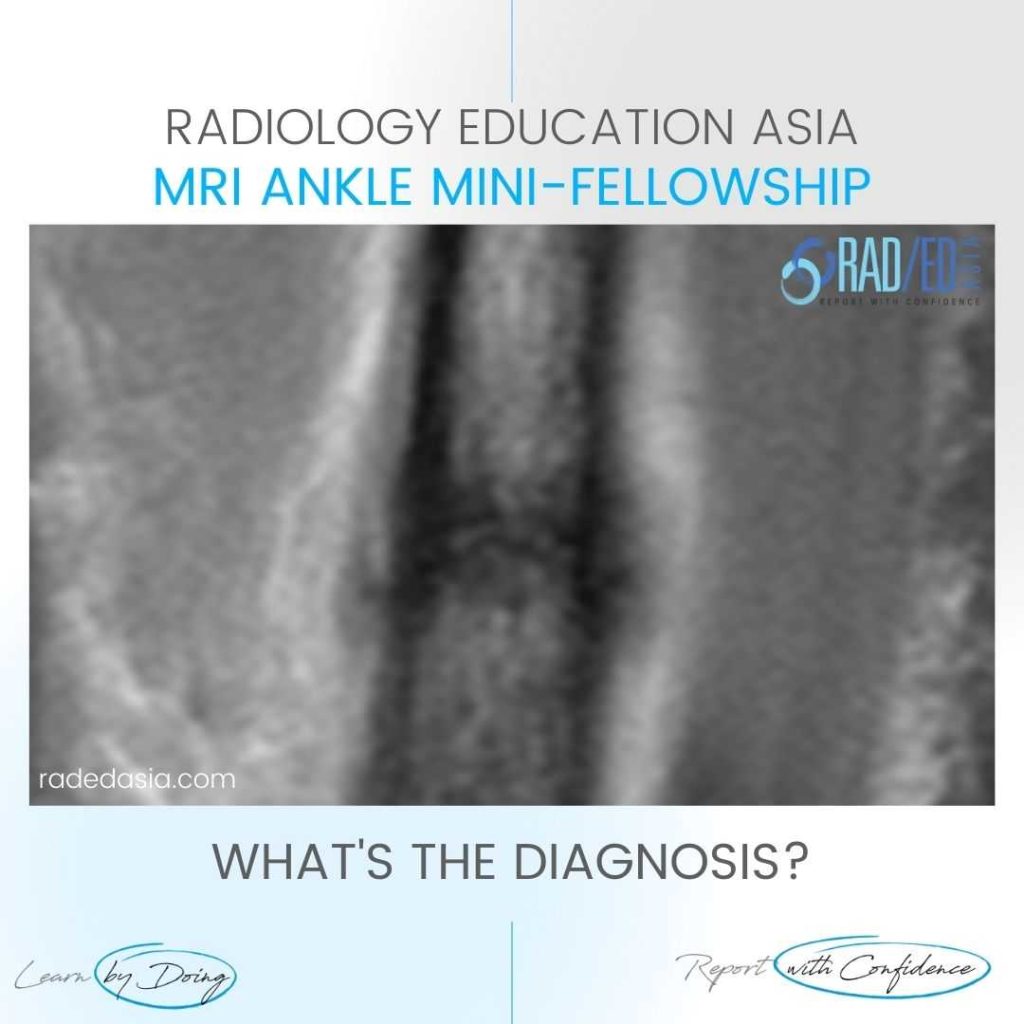
ANKLE

LEARN MSK MRI ANKLE ONLINE

ANKLE MSK MRI RADIOLOGY: We regularly post on Ankle MRI and our Ankle MRI Courses with all the posts on this page.

Ankle MRI can be difficult due to the complex anatomy. In our posts we have concise explanation of the Ankle MRI anatomy, the macroscopic pathology that’s relevant to the Imaging and the Imaging findings in various pathologies.

Learn where to Look, What to Look for and How to Report Ankle MRI More Confidently

msk ankle foot hip radiology workshop conference course musculoskeletal mri imaging kuala lumpur malaysia radedasia

MSK RADIOLOGY CONFERENCE COURSE MUSCULOSKELETAL MRI ANKLE FOOT & HIP MALAYSIA KUALA LUMPUR

Home MSK RADIOLOGY CONFERENCE COURSE MUSCULOSKELETAL MRI MALAYSIA KUALA LUMPUR: MSK MRI RADIOLOGY ANKLE, FOOT & HIP MINI FELLOWSHIP: 11/12 FEBRUARY 2023 This Mini Fellowship is now over.To see our latest Onsite Guided Mini Fellowships please click HERE WHAT HAPPENED AT THE MINI-FELLOWSHIP @ KL CLICK ON THE IMAGE BELOW TO FIND OUT Our 2-Day …

MSK RADIOLOGY CONFERENCE COURSE MUSCULOSKELETAL MRI ANKLE FOOT & HIP MALAYSIA KUALA LUMPUR Leer más »

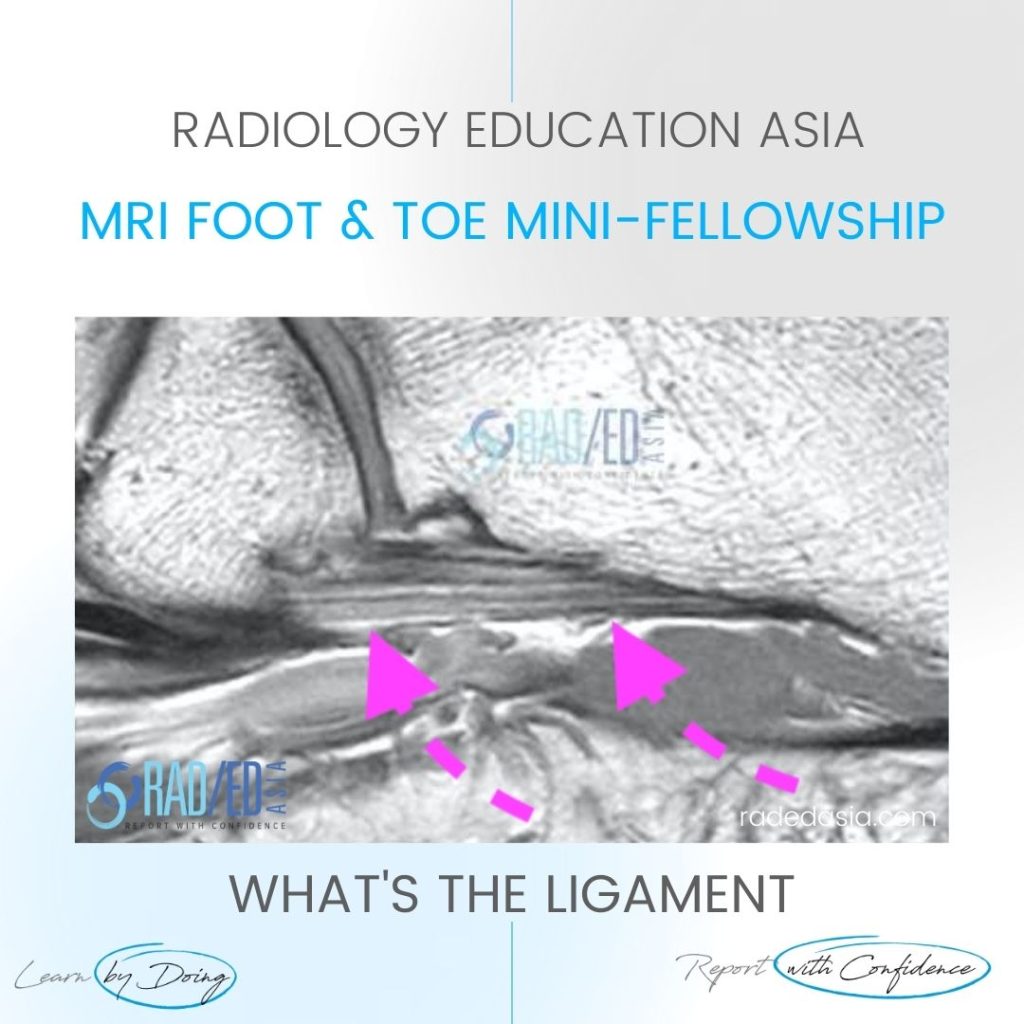
mri ankle foot ligament short plantar

MRI PLANTAR LIGAMENT SHORT PLANTAR LIGAMENT ANKLE FOOT AND TOE (VIDEO)

MRI PLANTAR LIGAMENT SHORT PLANTAR LIGAMENT WHAT'S THE STRUCTURE The Short Plantar Ligament. WHAT ARE THE FINDINGS ANATOMY OF THE SHORT PLANTAR LIGAMENT. 1. Extends from the calcaneus (Green star) to the cuboid (Yellow star). 2. It is distal and separate to long plantar ligament. 3. Has a fan shaped appearance. MRI PLANTAR LIGAMENT SHORT …

MRI PLANTAR LIGAMENT SHORT PLANTAR LIGAMENT ANKLE FOOT AND TOE (VIDEO) Leer más »

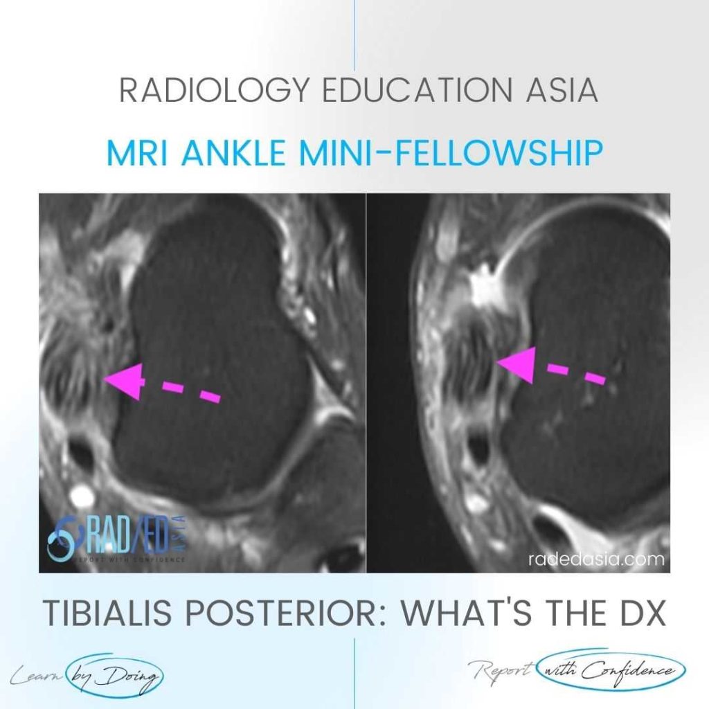
tibialis posterior tendinitis tendinopathy tear mri radiology

TIBIALIS POSTERIOR TENDINITIS TENDINOPATHY TEARS RADIOLOGY MRI (VIDEO)

TIBIALIS POSTERIOR TENDINITIS TENDINOPATHY TEARS RADIOLOGY MRI WHAT ARE THE FINDINGS The tibialis posterior tendon is enlarged and increased in signal. DIAGNOSIS & DISCUSSION TIBIALIS POSTERIOR TENDINOPATHY AND PARTIAL TEAR. Tibialis posterior tendinopathy and delamination / partial tears. The normal tibialis posterior tendon should be uniformly low signal. Enlargement and or increased intermediate signal indicates …

TIBIALIS POSTERIOR TENDINITIS TENDINOPATHY TEARS RADIOLOGY MRI (VIDEO) Leer más »

stress fracture fatigue insufficiency fracture radiology mri radedasia

STRESS FRACTURE FATIGUE FRACTURE AND INSUFFICIENCY FRACTURE WHAT IS THE DIFFERENCE (VIDEO)

FATIGUE FRACTURE AND INSUFFICIENCY FRACTURE: WHAT'S THE DIFFERENCE DISCUSSION The difference lies in the underlying bone metabolism. Is it normal or not. A Fatigue fracture is  stress type fracture due to repetitive overload, with normal bone metabolism (eg a March Fracture of the metatarsal in new army recruits). An Insufficiency fracture is a stress type …

STRESS FRACTURE FATIGUE FRACTURE AND INSUFFICIENCY FRACTURE WHAT IS THE DIFFERENCE (VIDEO) Leer más »

ankle impingement radiology anterior xray mri radedasia

ANKLE IMPINGEMENT RADIOLOGY ANTERIOR X-RAY (VIDEO)

ANKLE IMPINGEMENT RADIOLOGY ANTERIOR X-RAY DISCUSSION Ankle impingement can occur at multiple sites. One of the more common ones is Anterior.   The presence of Ankle impingement is ultimately a clinical diagnosis which is based on the patient’s symptoms and imaging findings, not just the imaging findings.   So in the report we describe the …

ANKLE IMPINGEMENT RADIOLOGY ANTERIOR X-RAY (VIDEO) Leer más »

mri plantar fascia plantar fasciitis plantar fascia tear learn radiology radedasia

MRI PLANTAR FASCIA TEAR vs PLANTAR FASCIITIS (VIDEO)

PLANTAR FASCIA TEAR vs PLANTAR FASCIITIS HOW TO DIFFERENTIATE BETWEEN PLANTAR FASCIA TEAR vs PLANTAR FASCIITIS:   How do you tell the difference on MRI between Plantar Fasciitis and Plantar Fascia Tears. Short 1 minute video on the key differences that help you differentiate a Plantar Fascia Tear from Plantar Fasciitis on MRI. VIEW VIDEO …

MRI PLANTAR FASCIA TEAR vs PLANTAR FASCIITIS (VIDEO) Leer más »

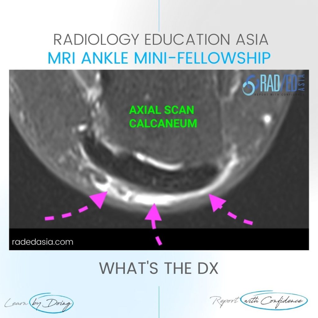
retroachilles bursa bursitis mri radiology ankle

RETROACHILLES BURSA BURSITIS MRI RADIOLOGY ANKLE

RETROACHILLES BURSA BURSITIS MRI ANKLE DISCUSSION ABOUT THE RETROACHILLES BURSA The retroachilles bursa lies between skin and Achilles tendon.   It’s not usually seen as its an adventitial bursa caused by chronic friction.   It is Uncommonly enlarged. WHERE TO LOOK FOR THE RETROACHILLES BURSA Best seen on axial and sagittal scans.   Look posterior …

RETROACHILLES BURSA BURSITIS MRI RADIOLOGY ANKLE Leer más »

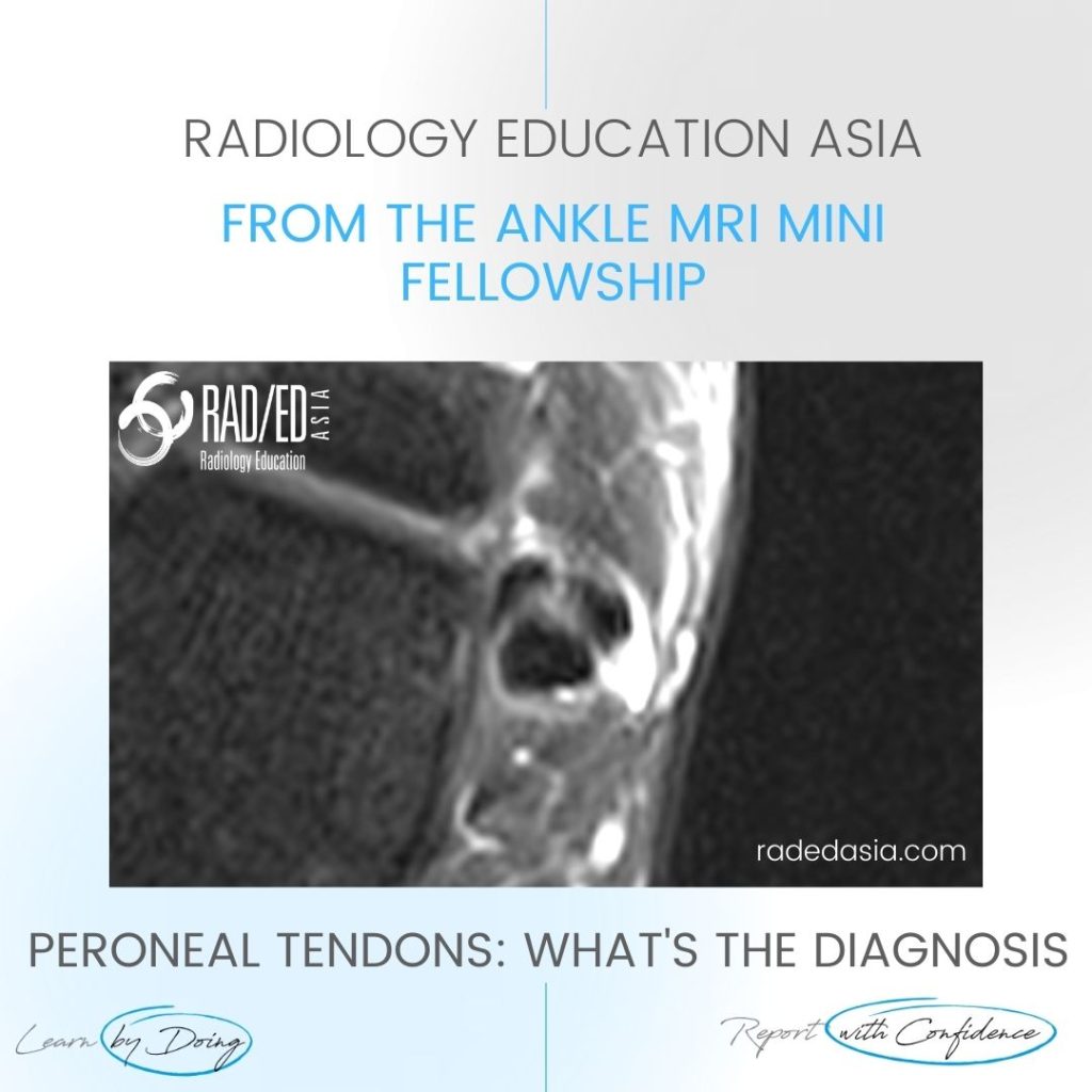
ankle mri peroneus brevis tendon tear split

PERONEUS BREVIS TENDON TEAR MRI ANKLE MRI

PERONEUS BREVIS TENDON TEAR MRI DISCUSSION: WHAT'S THE dX Peroneus Brevis Tendon Tear: MRI demonstrates a peroneus brevis tendon tear.  There is a longitudinal split resulting in two components of the Peroneus Brevis tendon (Pink arrows). Peroneus Longus (Blue arrow) is normal. WHAT ARE THE FINDINGS There should only be two tendons in the peroneal …

PERONEUS BREVIS TENDON TEAR MRI ANKLE MRI Leer más »

Este sitio está destinado únicamente a profesionales médicos. El uso de este sitio se rige por nuestra Términos de servicio y Declaracion de privacidad que se puede encontrar haciendo clic en los enlaces. Acepte antes de continuar con el sitio web.
error: Clic derecho no disponible.