Spine MRI Mini-Fellowship and Workstation Workshop Singapore
CLICK HERE FOR OUR LATEST SPINE MRI MINI-FELLOWSHIPS
CLICK HERE FOR OUR LATEST SPINE MRI MINI-FELLOWSHIPS
The Problem: Acute cerebellar infarcts are easy to see on diffusion imaging, but small chronic infarcts in the cerebellum are often missed because they are confused with normal cerebellar sulci. How do you differentiate small chronic cerebellar infracts from normal sulci? The Answer: In the Cerebellar Hemispheres: Normal: Chronic Infarct: Infarcts usually run perpendicular to the …
Registrations have now closed for the MSK MRI Workstation based workshop to be held in Kuala Lumpur Malaysia in July 2016. We would like to thank everyone who registered and to those who expressed interest. Due to continuing interest in the workshop we have decided to hold a second workshop in KL on the weekend …
MSK MRI Workstation Workshop KUALA LUMPUR MALAYSIA Read More »

CHONDRAL DELAMINATION CARTILAGE MRI CHONDRAL DELAMINATION CARTILAGE MRI Chondral Delamination on MRI is not common but is seen often enough that we need to be aware of it and What to look for on MRI. OVERVIEW What is it? Chondral delamination is when cartilage separates and lifts off from its attachment with the cortex. …
CHONDRAL DELAMINATION CARTILAGE MRI: Quick Review Read More »

When scans are performed for ? Discitis, often both a T2 and T2 Fat Sat/ STIR sequence are performed. Which is better for assessing for early discitis? One of the features of discitis on MRI is increased T2 signal in the disc. But often, there are no other features of discitis/ osteomyelits, the disc signal …
The first MSK MRI Workstation based workshop in Kuala Lumpur is now confirmed for the weekend of the 23rd and 24th of July 2016. We will cover MRI of the four most commonly imaged joints, Knee, Shoulder, Hip and Ankle. The best way to learn something is to do it. Lectures and conferences give you …
MSK MRI WORKSTATION WORKSHOP: KUALA LUMPUR 23-24TH JULY Read More »
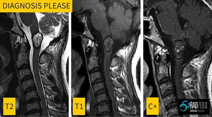
This is the first in a regular series of posts on cases of interest. Incidental finding in a 40yo male. Image above. ANSWER Cavernomas of the spinal cord have a very specific appearance on MRI with the following features Lobulated well defined lesion mixed signal T1 and T2 No significant enhancement Low signal …
Radiology Education Diagnosis Please: Spinal cord lesion MRI Read More »

Going to Bali or Borocay for a holiday and just have to read all the radiology articles in AJR and Radiographics you missed in the year? Work at different sites during the week or have limited internet access? How do you get easy access to articles? I carry around a few thousand articles in my …
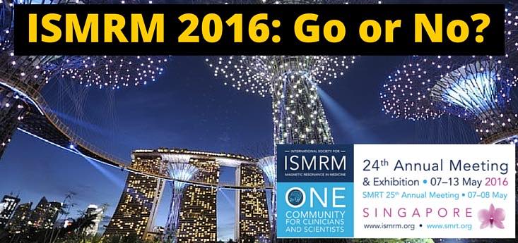
The 2016 ISMRM Annual Meeting is being held in Singapore, which is great news for us in this region. But is it worth going to? It depends…. I have been to a number of ISMRM meetings over the years and its a huge meeting, but the difficulty from a radiologist’s perspective, is that it …

In the last post we looked at the Buford Complex. In this post lets look at the remaining two normal labral variants, the Sublabral Recess and the Sublabral Foramen Definition: Regions where a normal labrum is present but it is not attached to the glenoid. Where are they found? Sublabral Recess: Anterior portion of the …
GLENOID LABRUM MRI SIMPLIFIED 4: Sublabral Foramen and Recess Read More »