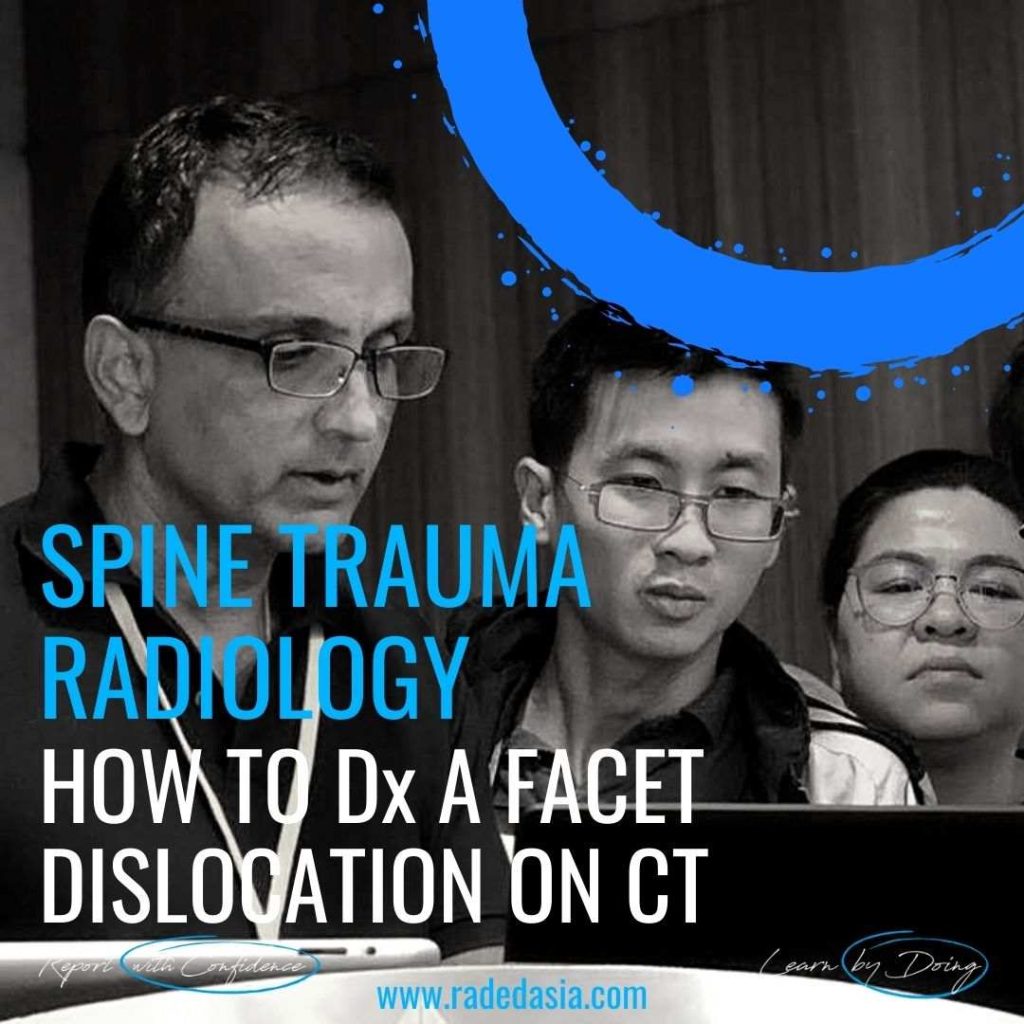
SPINE TRAUMA RADIOLOGY: HOW TO Dx A FACET DISLOCATION ON CT (VIDEO)
SPINE TRAUMA RADIOLOGY: FACET DISLOCATION ON CT FACET DISLOCATION ON CT: Spinal trauma can result in a number of abnormalities of the facet joints ranging from Diastasis, Subluxation, Perched Facets and eventually to Dislocation or Locked Facets. In this short video we look at the normal appearance of the facet joints, how that …
SPINE TRAUMA RADIOLOGY: HOW TO Dx A FACET DISLOCATION ON CT (VIDEO) Read More »