RHEUMATOLOGY

Arthritis Radiology Rheumatology Imaging

Imaging of Arthritis can be challenging because findings can be non specific and the clinical context of what we are seeing is very important in getting to or narrowing a diagnosis.

From 2021 all our posts on Arthritis Radiology and Rheumatology Imaging will be done in conjunction with Dr Joe Thomas a Senior Consultant Rheumatologist. who has a vast amount of clinical experience.

He also has a very strong interest in Imaging of Arthropathies and has joined us to bring a clinical perspective to the imaging and to advise on what rheumatologists want when we report their referrals.

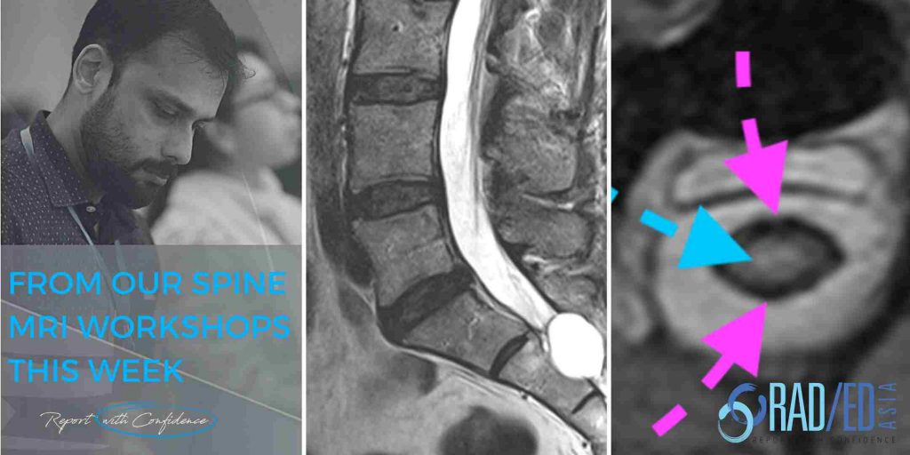
schmorl nodes spine mri online radiology courses

SCHMORL’S NODES SPINE MRI ONLINE RADIOLOGY COURSE LEARN KEY POINTS

KEY POINTS FROM OUR SPINE MRI COURSES Schmorl’s Node Spine MRI Online Radiology Course Acute and Chronic Schmorl’s nodes. Another three quick images with some basic key points from our online spine MRI courses. ACUTE SCHMORL'S NODE MRI Acute schmorl’s nodes demonstrate oedema at their margins. This is usually limited to around the circumference of …

SCHMORL’S NODES SPINE MRI ONLINE RADIOLOGY COURSE LEARN KEY POINTS Read More »

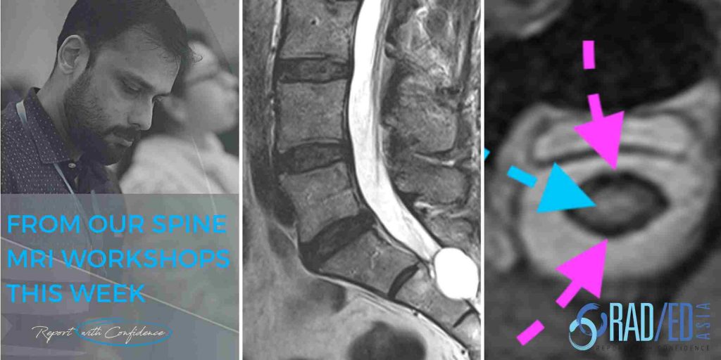
arachnoiditis spine mri online radiology courses radedasia

ARACHNOIDITIS MRI SPINE ONLINE RADIOLOGY COURSE

KEY POINTS FROM OUR SPINE MRI COURSES Arachnoiditis Spine MRI Online Radiology Course What are some of the common appearance of arachnoiditis on MRI? Another three quick images with some basic key points from our online spine MRI courses. ARACHNOIDITIS MRI ASYMMETRIC DISTRIBUTION WHAT'S THE SIGNIFICANCE OF ASYMMETRY? The distribution of nerve roots in the …

ARACHNOIDITIS MRI SPINE ONLINE RADIOLOGY COURSE Read More »

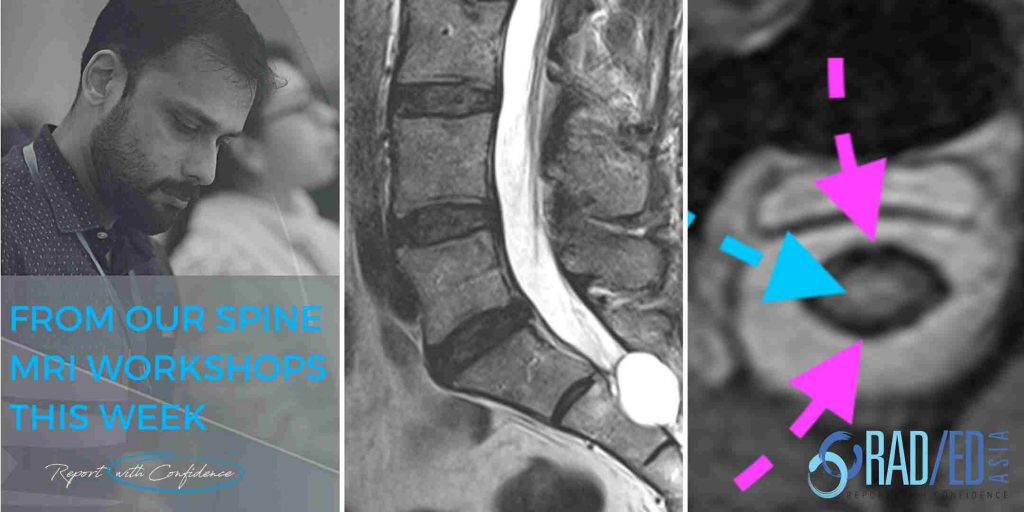
opll Ossification Posterior Longitudinal Ligament X-ray, CT, MRI spine mri online radiology course

OPLL SPINE OSSIFICATION POSTERIOR LONGITUDINAL LIGAMENT CERVICAL

OPLL SPINE OSSIFICATION POSTERIOR LONGITUDINAL LIGAMENT CERVICAL OPLL SPINE Ossification Posterior Longitudinal Ligament Cervical Spine: X-ray, CT, MRI What does OPLL of the Cervical Spine  look like on MRI, CT and X-ray? Another three quick images with some basic key points from our online spine MRI courses. How to report OPLL X-Ray. How to report …

OPLL SPINE OSSIFICATION POSTERIOR LONGITUDINAL LIGAMENT CERVICAL Read More »

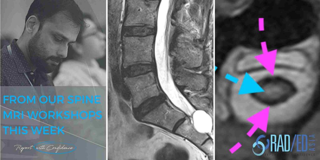
spine facet joint mri online radiology courses

SPINE FACET JOINT MRI ONLINE RADIOLOGY COURSES: KEY POINTS FROM OUR SPINE MRI COURSES

KEY POINTS FROM OUR SPINE MRI COURSES Spine Facet Joint MRI Online Radiology Course Facet joints can have a number of pathologies affecting them. Here we have three quick images with some key points about facet joint degeneration, facet joint ankylosis and facet joint infection from our online spine mri courses. SPINE MRI FACET JOINT …

SPINE FACET JOINT MRI ONLINE RADIOLOGY COURSES: KEY POINTS FROM OUR SPINE MRI COURSES Read More »

spine mri msk online radiology courses radedasia

SPINE MRI ONLINE RADIOLOGY COURSE NERVE ROOTS : KEY POINTS FROM OUR SPINE MRI COURSES

KEY POINTS FROM OUR SPINE MRI COURSES Spine MRI Online Radiology Course: How do you describe the nerve roots in your report.  Another three quick images with key points from our online spine mri courses on the basic nerve root nomenclature to use when describing which nerve is compressed.  Here we look at the appearance …

SPINE MRI ONLINE RADIOLOGY COURSE NERVE ROOTS : KEY POINTS FROM OUR SPINE MRI COURSES Read More »

ankylosing spondylitis spine imaging dagger sign trolley track sign spine fusion xray ct mri

ANKYLOSING SPONDYLITIS DAGGER SIGN SPINE IMAGING : WHAT DOES IT MEAN

ANKYLOSING SPONDYLITIS DAGGER SIGN SPINE IMAGING In ankylosing spondylitis, what is the dagger sign? In this post we look at  how to recognise it and  he Anatomy and Pathology that helps explain what the Dagger sign is. WHAT IS THE DAGGER SIGN In Ankylosing spondylitis the dagger sign refers to a solid central line of …

ANKYLOSING SPONDYLITIS DAGGER SIGN SPINE IMAGING : WHAT DOES IT MEAN Read More »

ankylosing spondylitis hip involvement imaging radedasia

ANKYLOSING SPONDYLITIS HIP PERIPHERAL JOINT INVOLVEMENT

ANKYLOSING SPONDYLITIS HIP PERIPHERAL JOINT INVOLVEMENT RADIOLOGY HOW COMMON IS HIP JOINT INVOLVEMENT IN ANK SPOND: Hip involvement in Ankylosing spondylitis has been reported in up to a 1/3rd of patients and is usually bilateral. WHY DOES IT OCCUR: Its primarily an inflammatory process. This results in cartilage destruction and loss of joint space. Eventually …

ANKYLOSING SPONDYLITIS HIP PERIPHERAL JOINT INVOLVEMENT Read More »

romanus lesion mri radiology education asia

ROMANUS LESION MRI FINDINGS CORNER INFLAMMATORY LESIONS

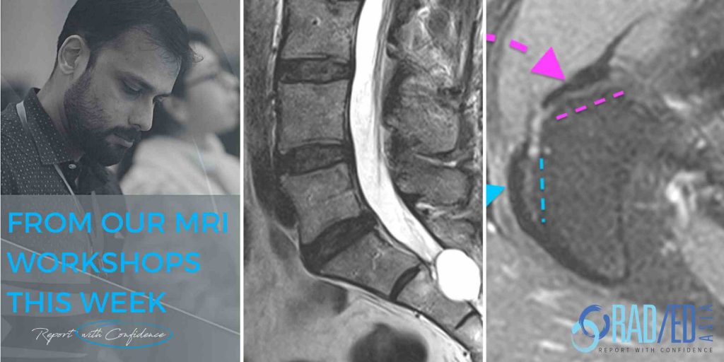
ROMANUS LESION MRI CORNER INFLAMMATORY LESIONS ROMANUS LESION CORNER INFLAMMATORY LESION MRI   Romanus Lesions are now called Corner Inflammatory Lesions. This post is the last in the series on Romanus lesions in ankylosing spondylitis and looks at the MRI findings in both acute and chronic changes. (If you haven’t seen the first two posts, …

ROMANUS LESION MRI FINDINGS CORNER INFLAMMATORY LESIONS Read More »

This site is intended for Medical Professionals only. Use of this site is governed by our Terms of Service and Privacy Statement which can be found by clicking on the links. Please accept before proceeding to the website.
error: Right Click not available.